
Ophthalmology
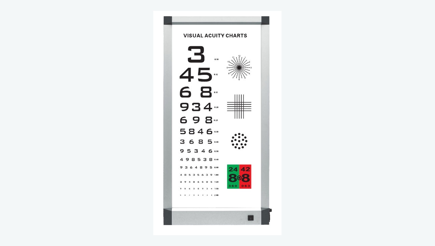
Visual Acuity Chart Light Box
Displays standardized distance, near or pediatric optotypes for consistent visual acuity testing.
Medical Equipment
Plan optometry, cataract and vitreoretinal equipment around examination pathways, the treatment scope and local clinical readiness.
Send a category enquiry or BOQOpen a reference to review its use and configuration guidance, or send an enquiry for that item. References retain their configuration identity so you can compare options without combining their quantities.
Eye clinics, hospital eye centres and specialist service development.
76 equipment references
Displays standardized distance, near or pediatric optotypes for consistent visual acuity testing.
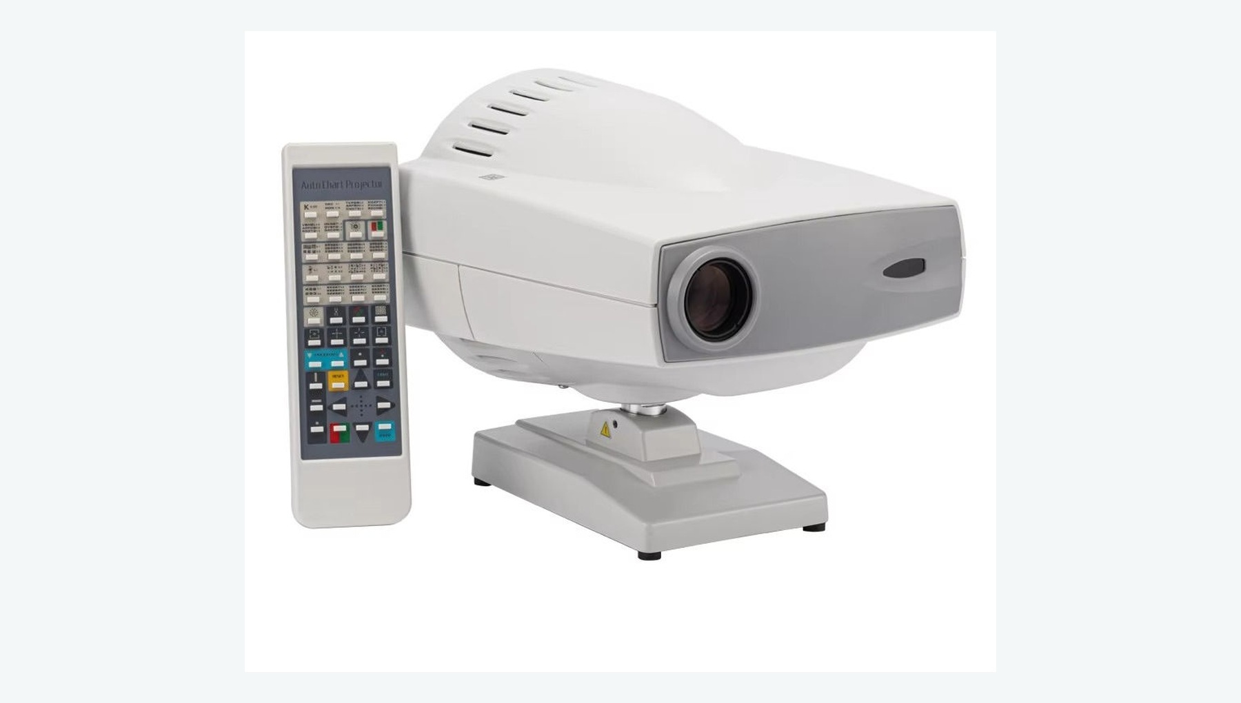
Projects standardized optotypes onto a screen for distance visual acuity assessment and a range of visual tests.
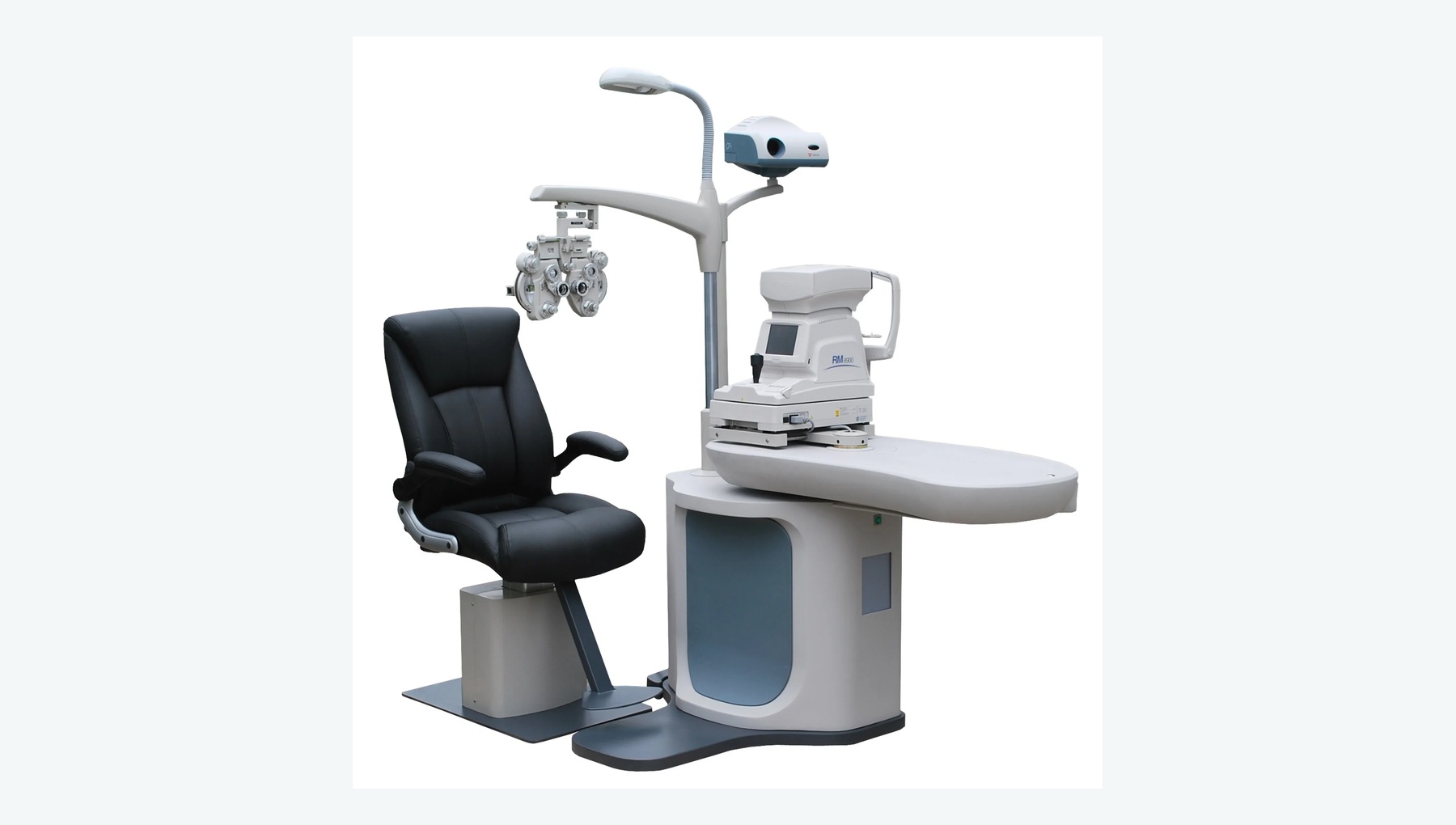
Combines the examination chair, adjustable instrument table and routine ophthalmic devices in a standardized refraction station.
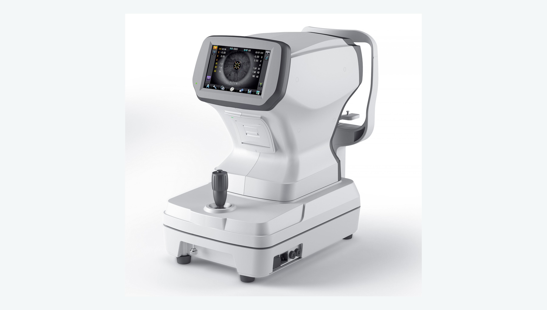
Rapidly measures objective refraction, cylinder axis and corneal curvature to inform subjective refraction.
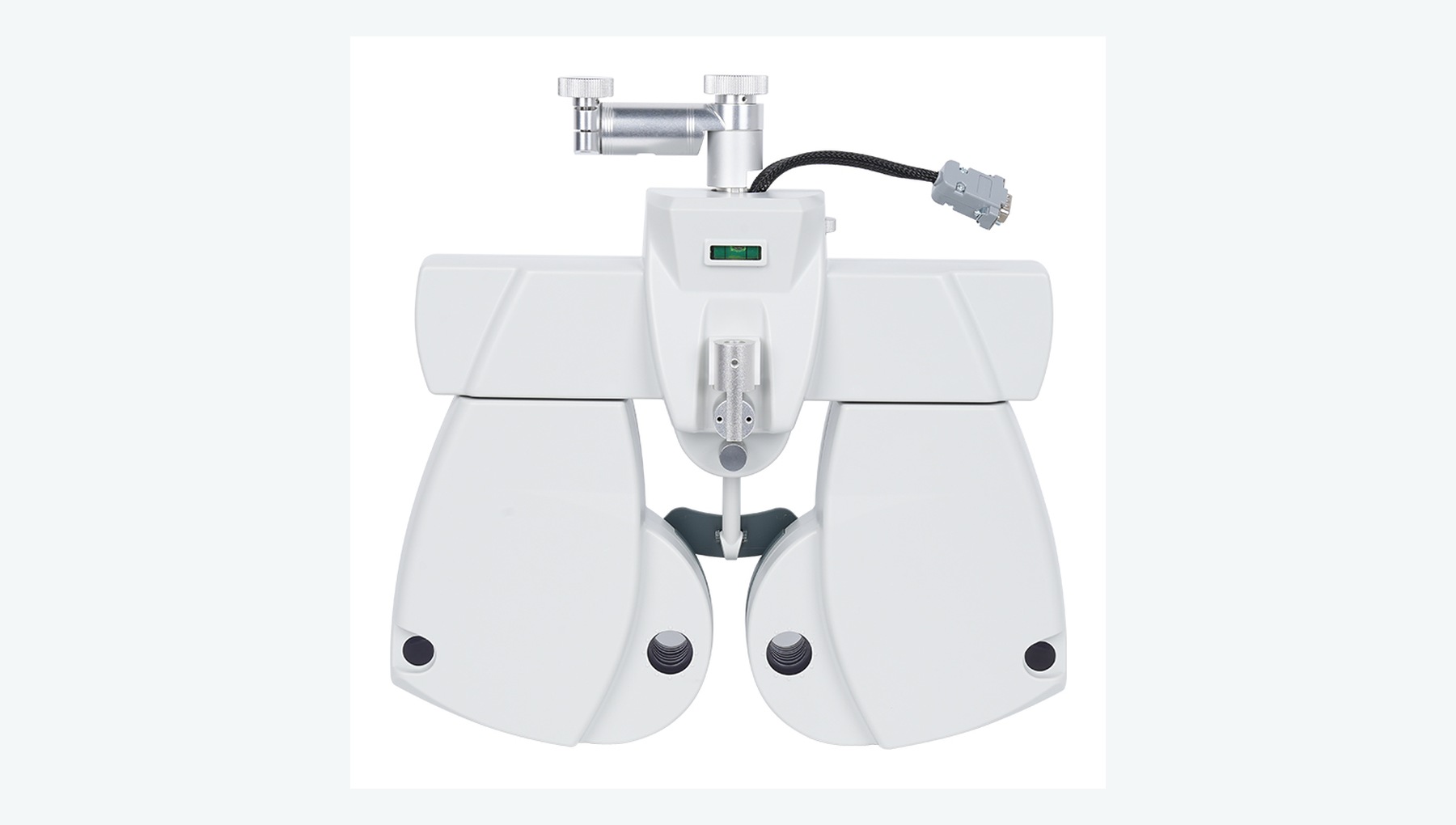
Supports refined subjective refraction using spherical and cylindrical lenses, Jackson cross-cylinder testing and binocular balance.
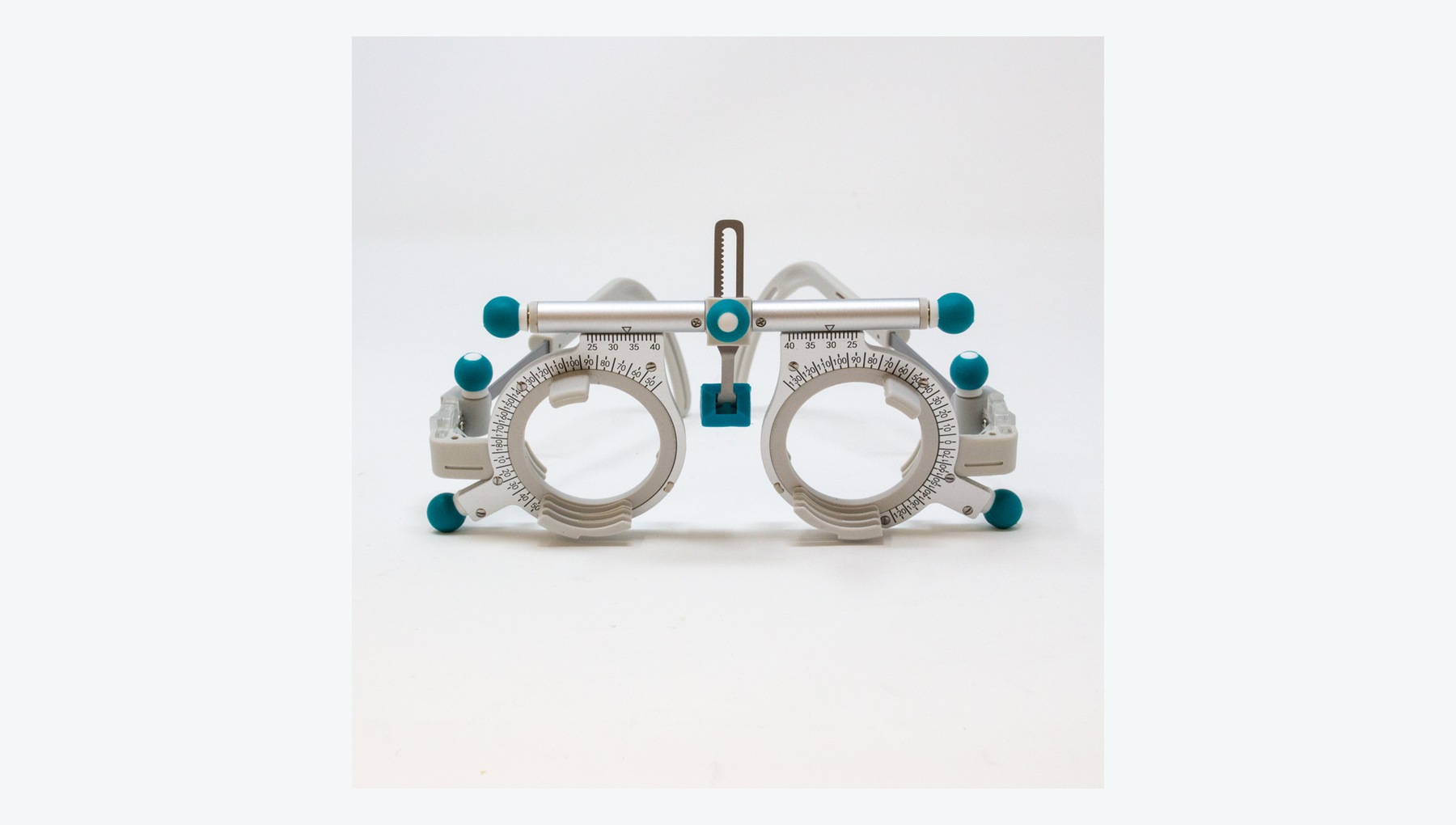
Holds spherical, cylindrical or prismatic trial lenses for subjective refraction, prescription verification and trial wear.
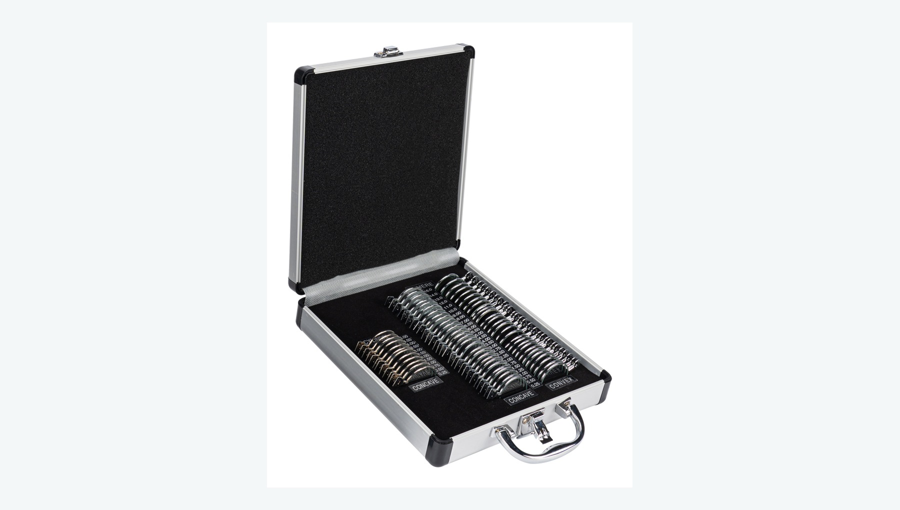
Provides spherical, cylindrical, prismatic and accessory lenses across a range of powers for manual refraction and prescription verification.
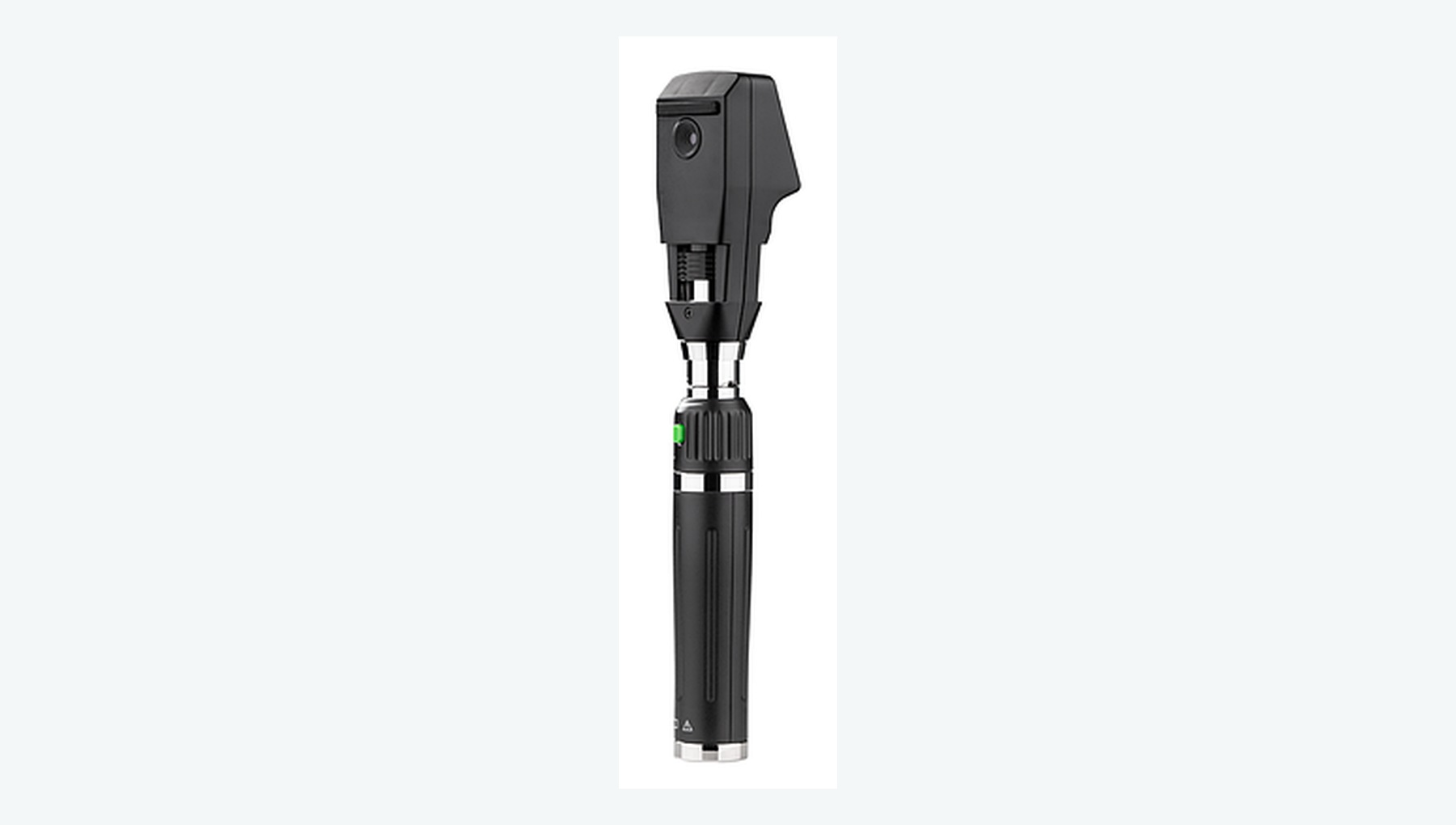
Provides objective refraction from the retinal reflex, particularly for children or patients with limited ability to cooperate.
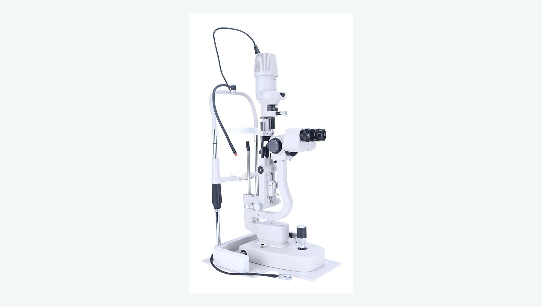
Provides magnified examination of the cornea, conjunctiva, anterior chamber, crystalline lens and ocular surface.
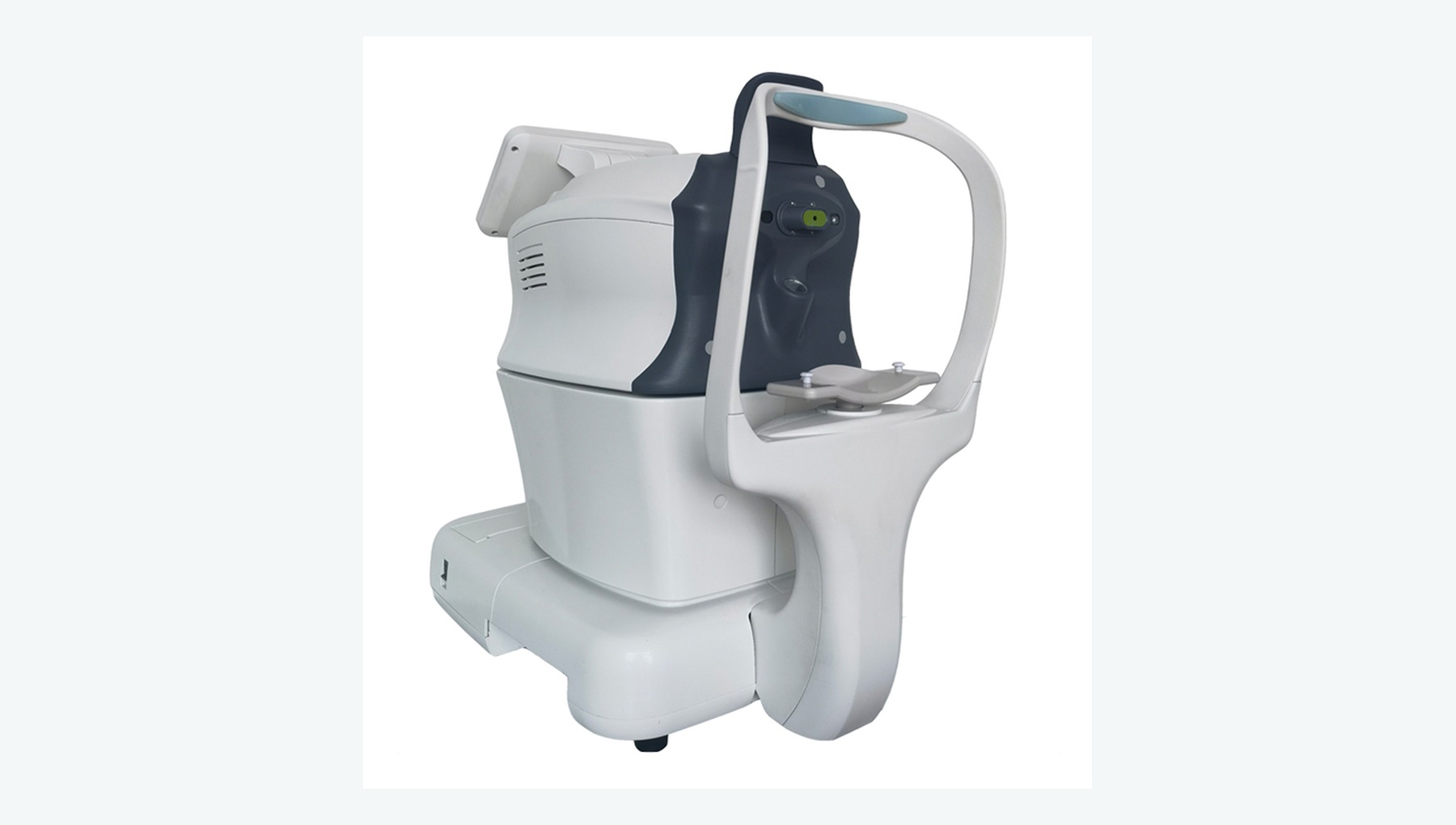
Measures intraocular pressure rapidly and helps identify patients with abnormal readings.
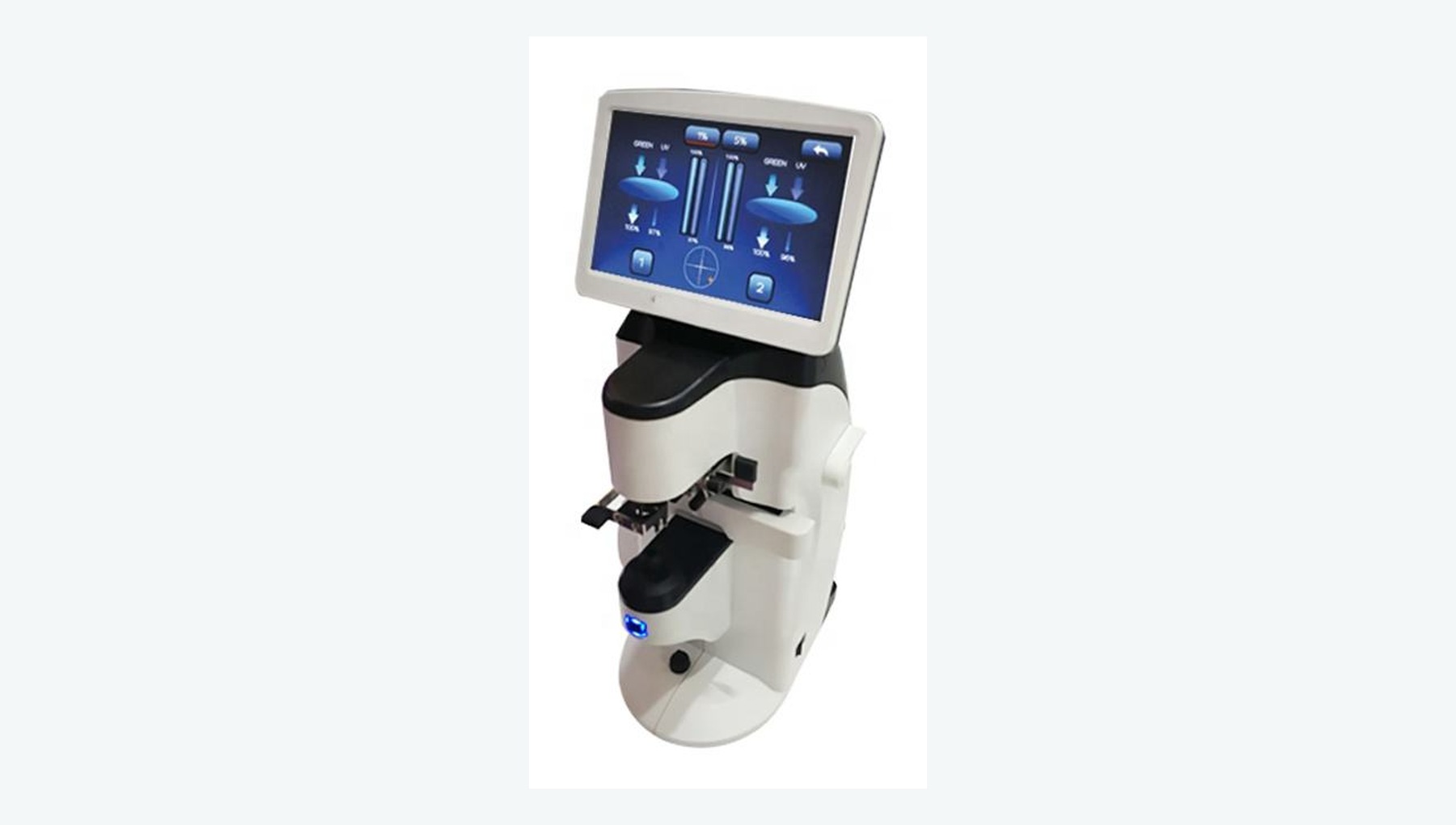
Measures sphere, cylinder, axis, prism and optical-center position in existing spectacles.
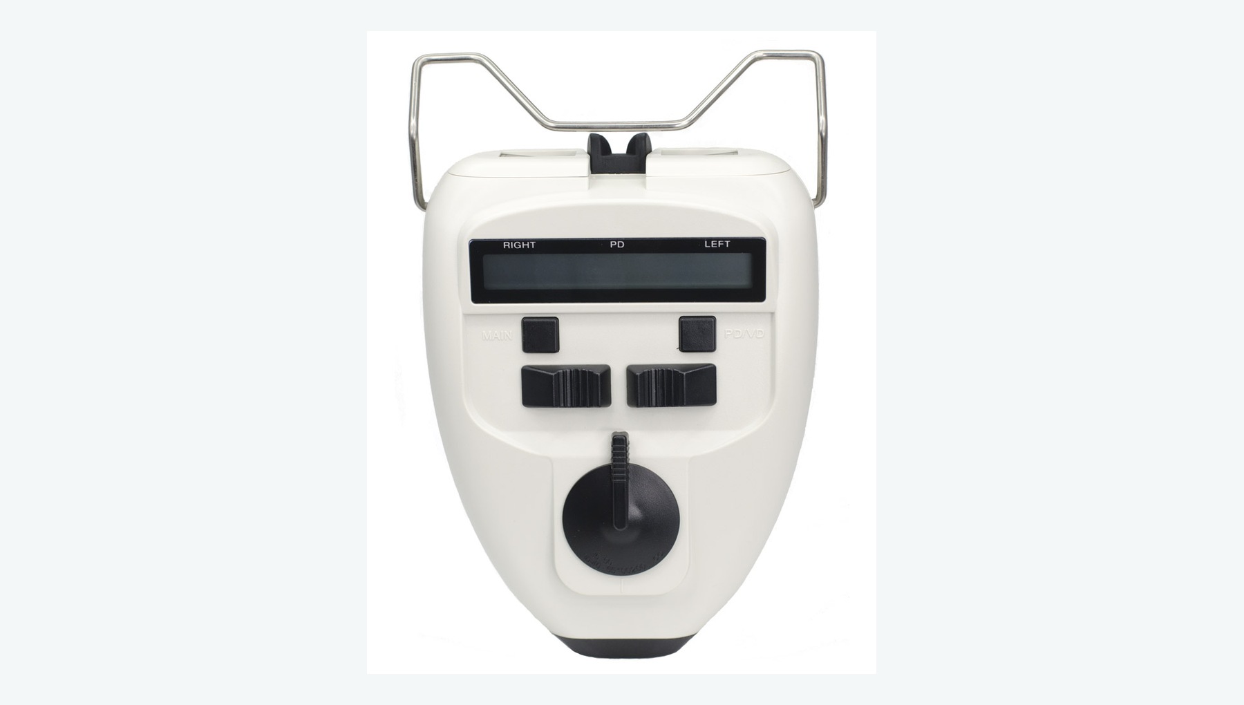
Measures monocular or binocular pupillary distance to support accurate optical-center positioning.
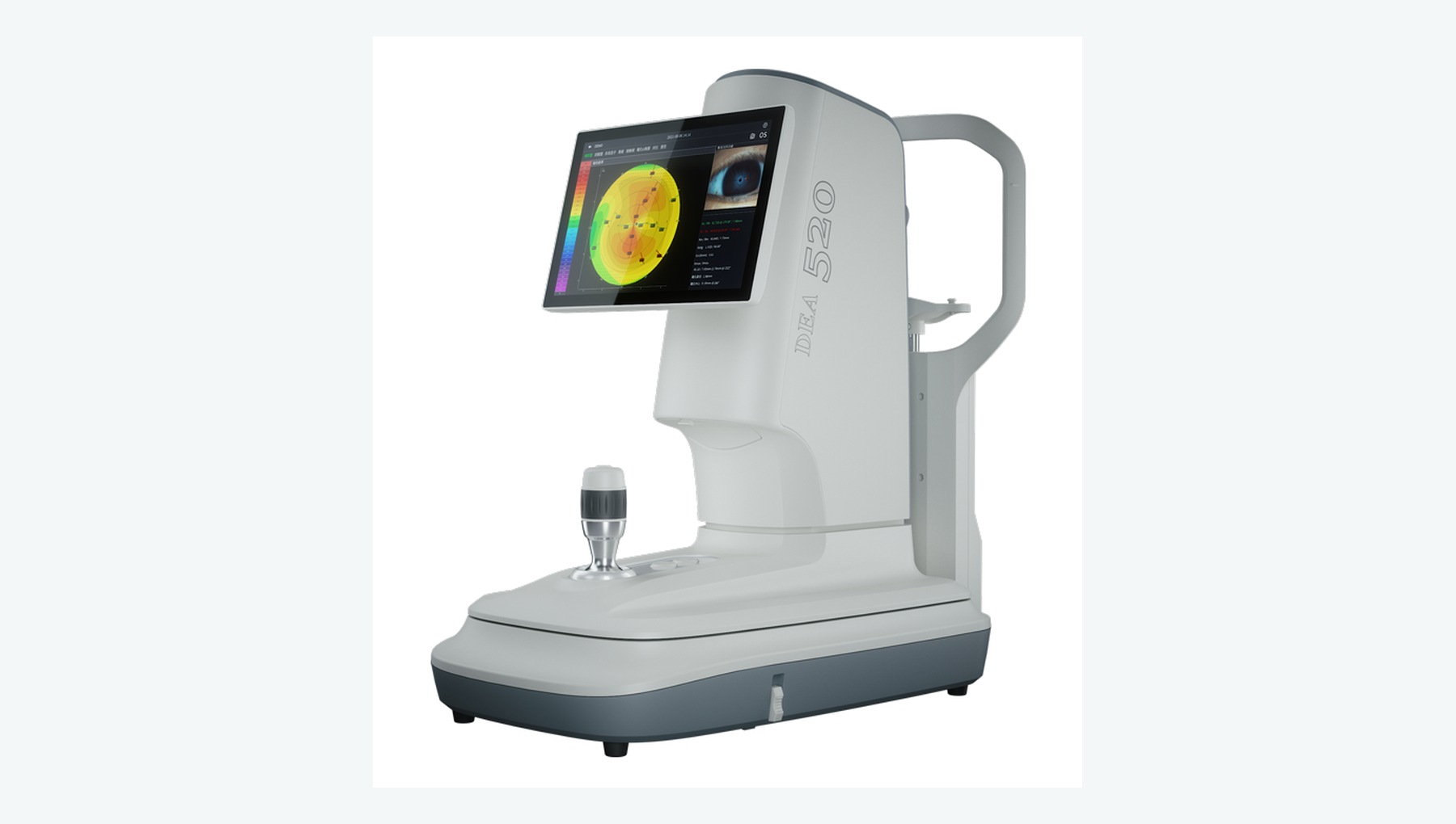
Analyzes corneal curvature, shape and regularity to support complex contact lens fitting.
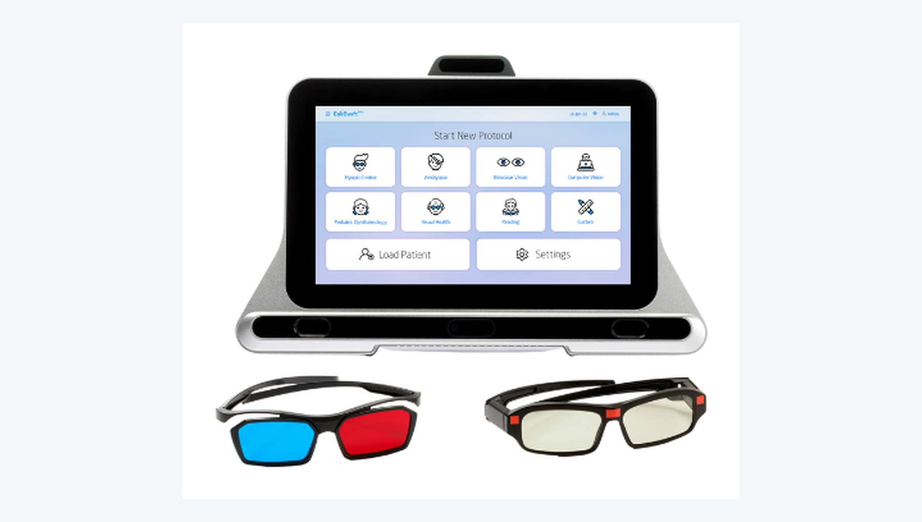
Assesses ocular alignment, accommodation, convergence, fusion and stereopsis, with support for basic vision training.
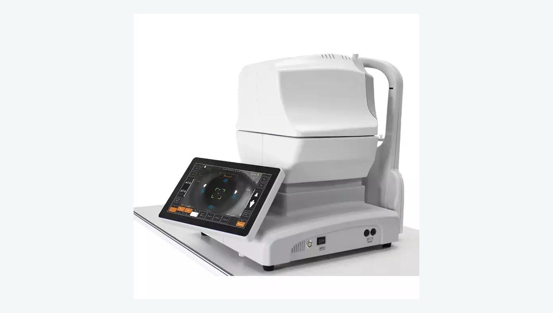
Captures retinal and optic-disc images to support initial fundus-disease screening and referral.
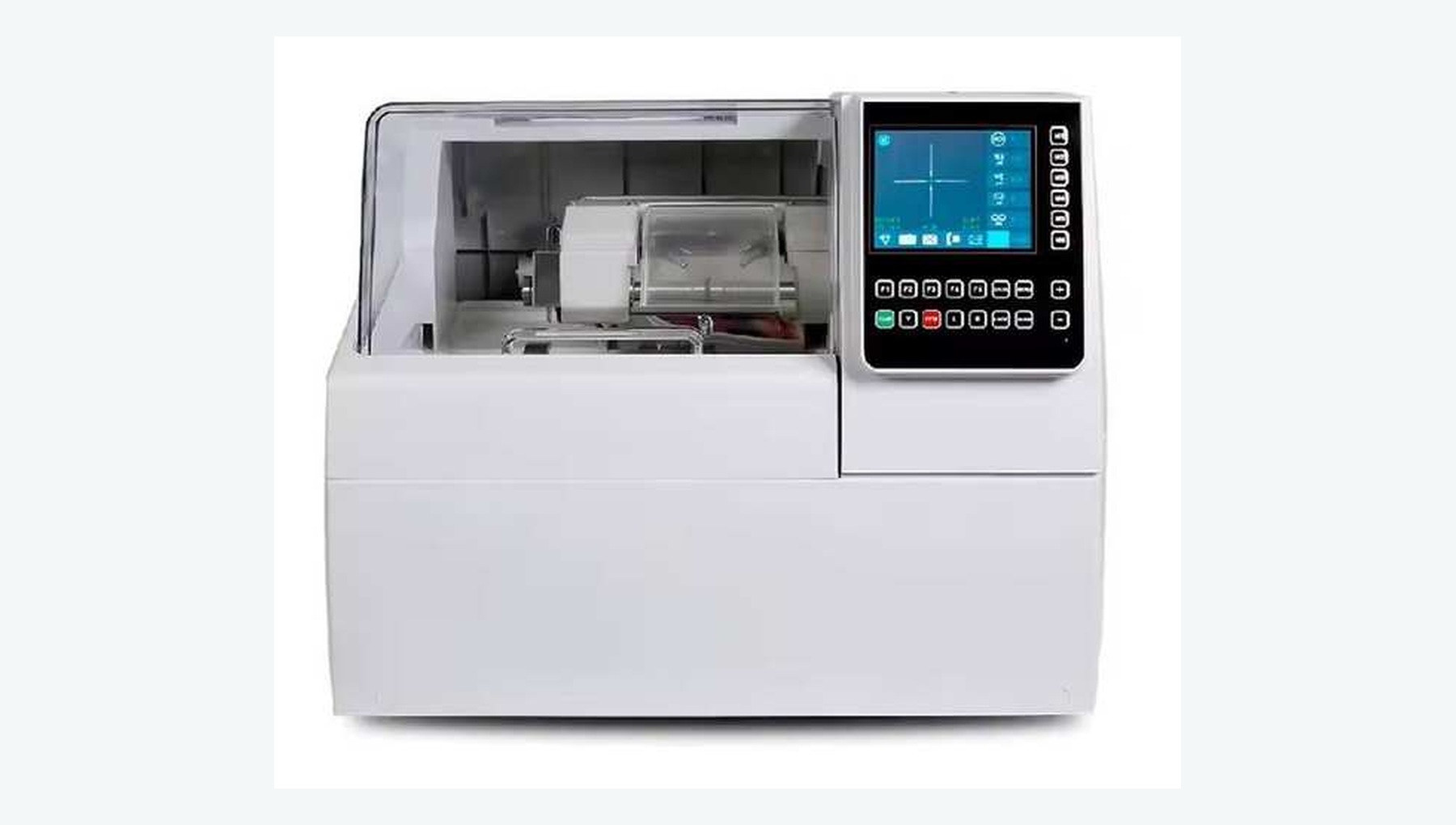
Handles lens tracing, centering, edging, beveling and assembly.
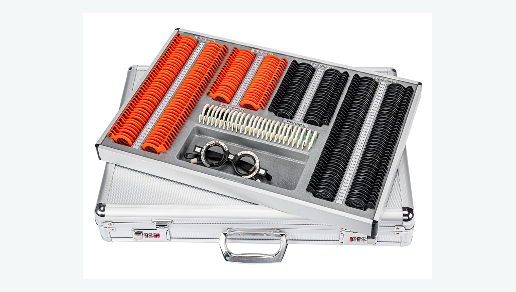
Brings together routine tools such as a trial frame, trial lenses, occluder, pupillary-distance ruler and near-vision chart.
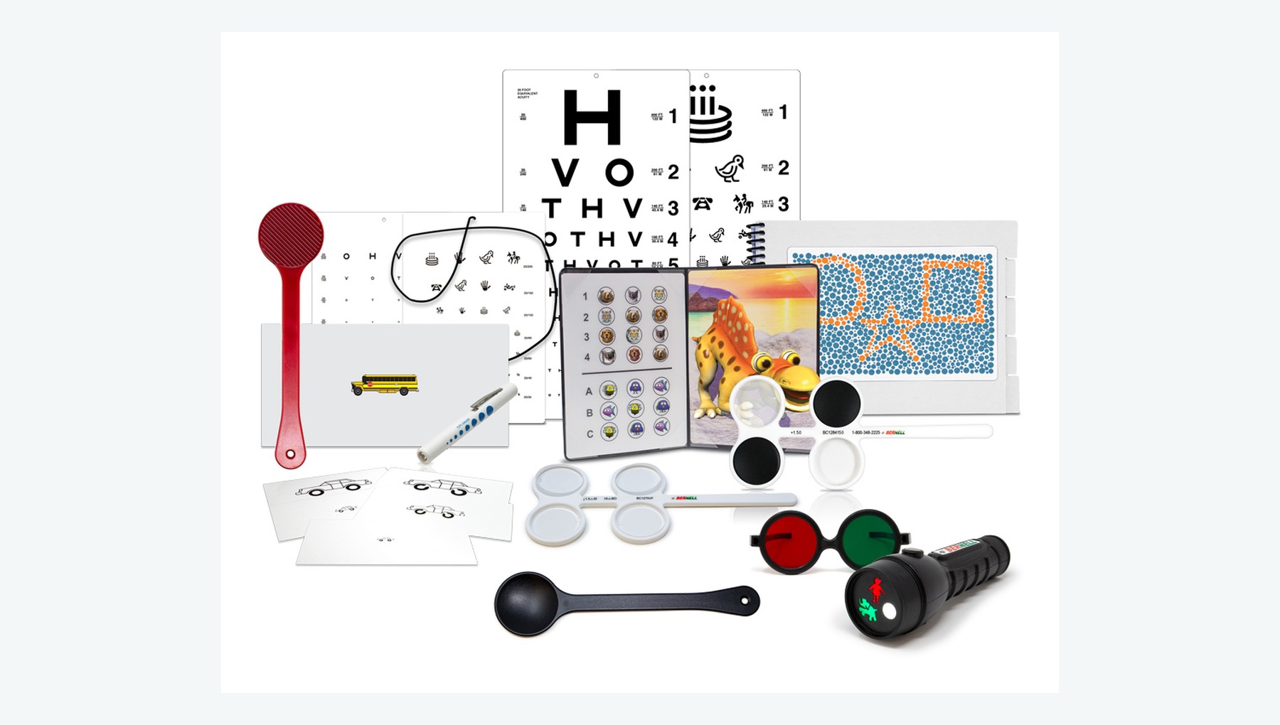
Supports assessment of visual acuity, ocular alignment, accommodation, convergence, color vision and stereopsis in children.
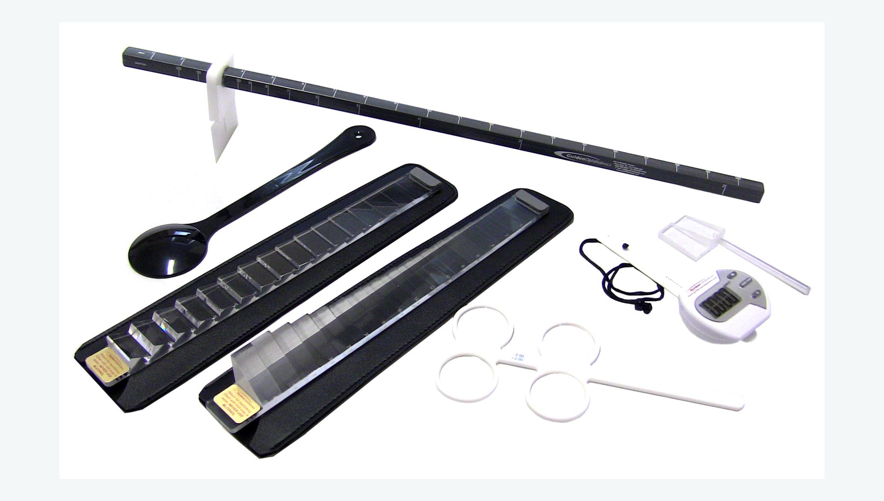
Supports assessment of heterophoria, fusional ranges, accommodation and stereopsis.
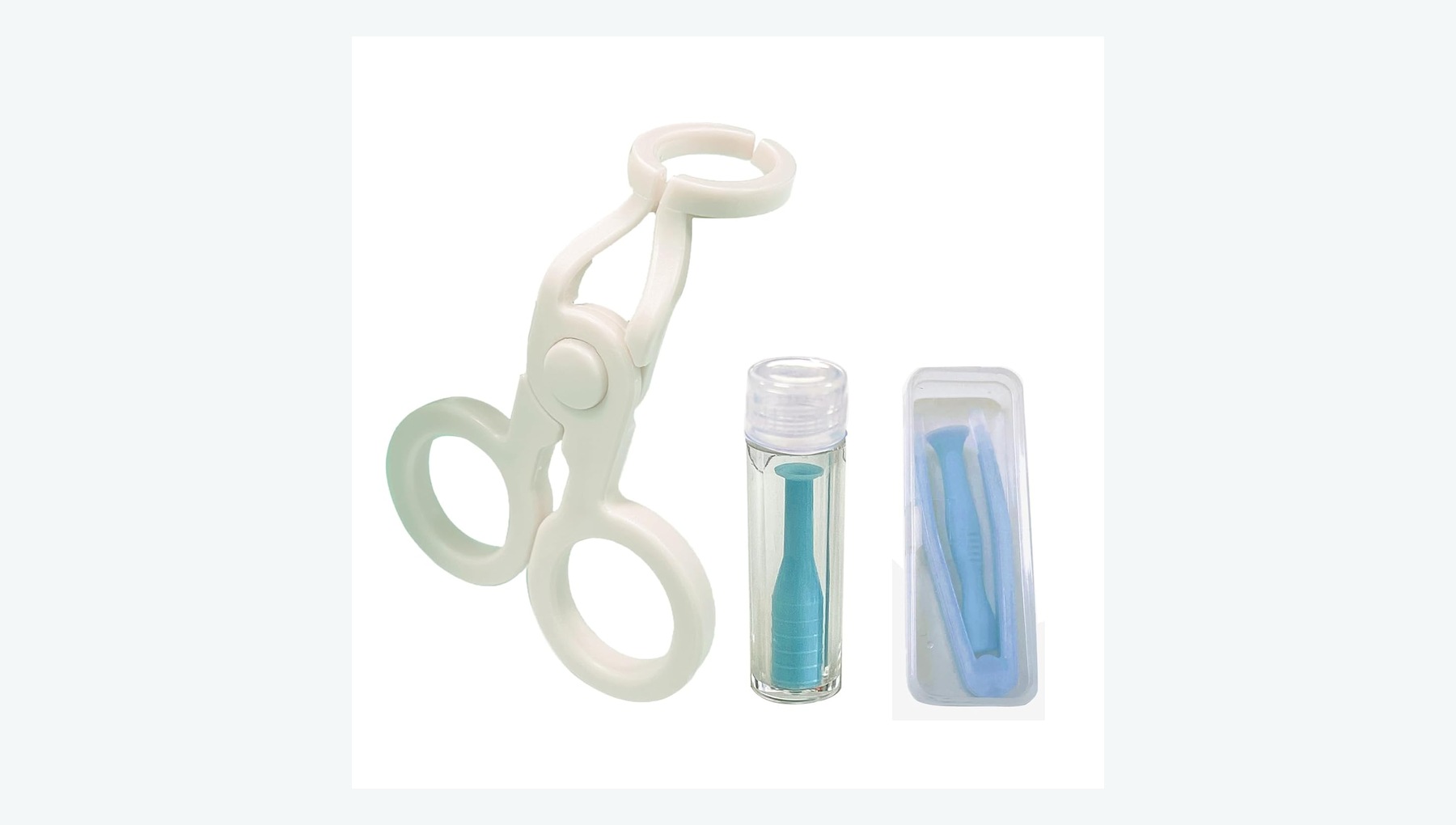
Supports contact lens trials, insertion and removal training, fluorescein assessment and lens care.
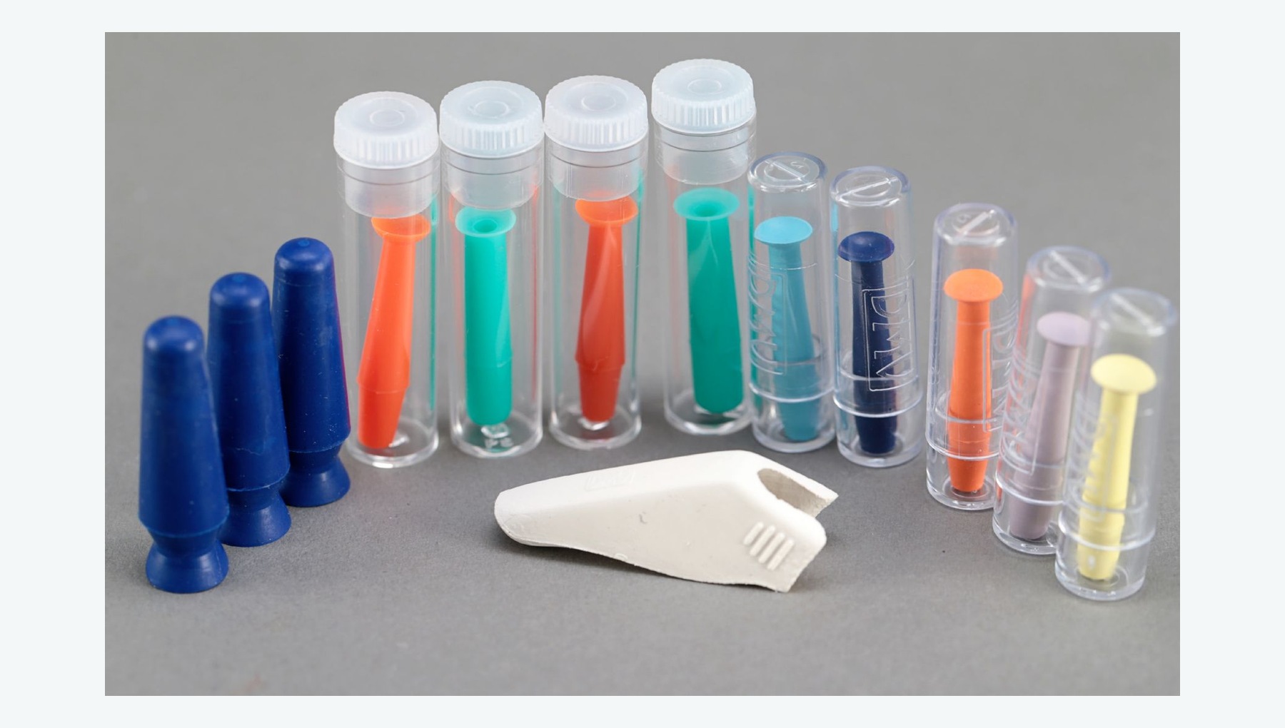
Supports trial fitting, fit assessment and follow-up for rigid gas-permeable and orthokeratology lenses.
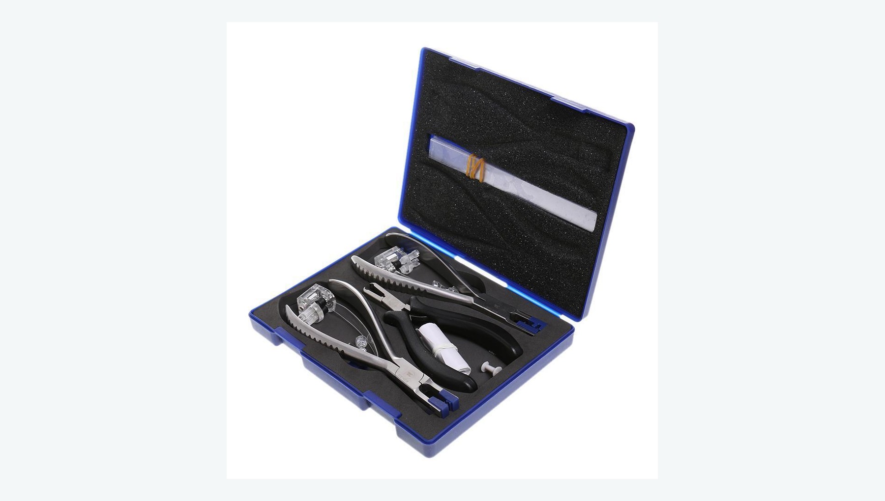
Supports frame adjustment, assembly, repair and optical quality checks on finished spectacles.
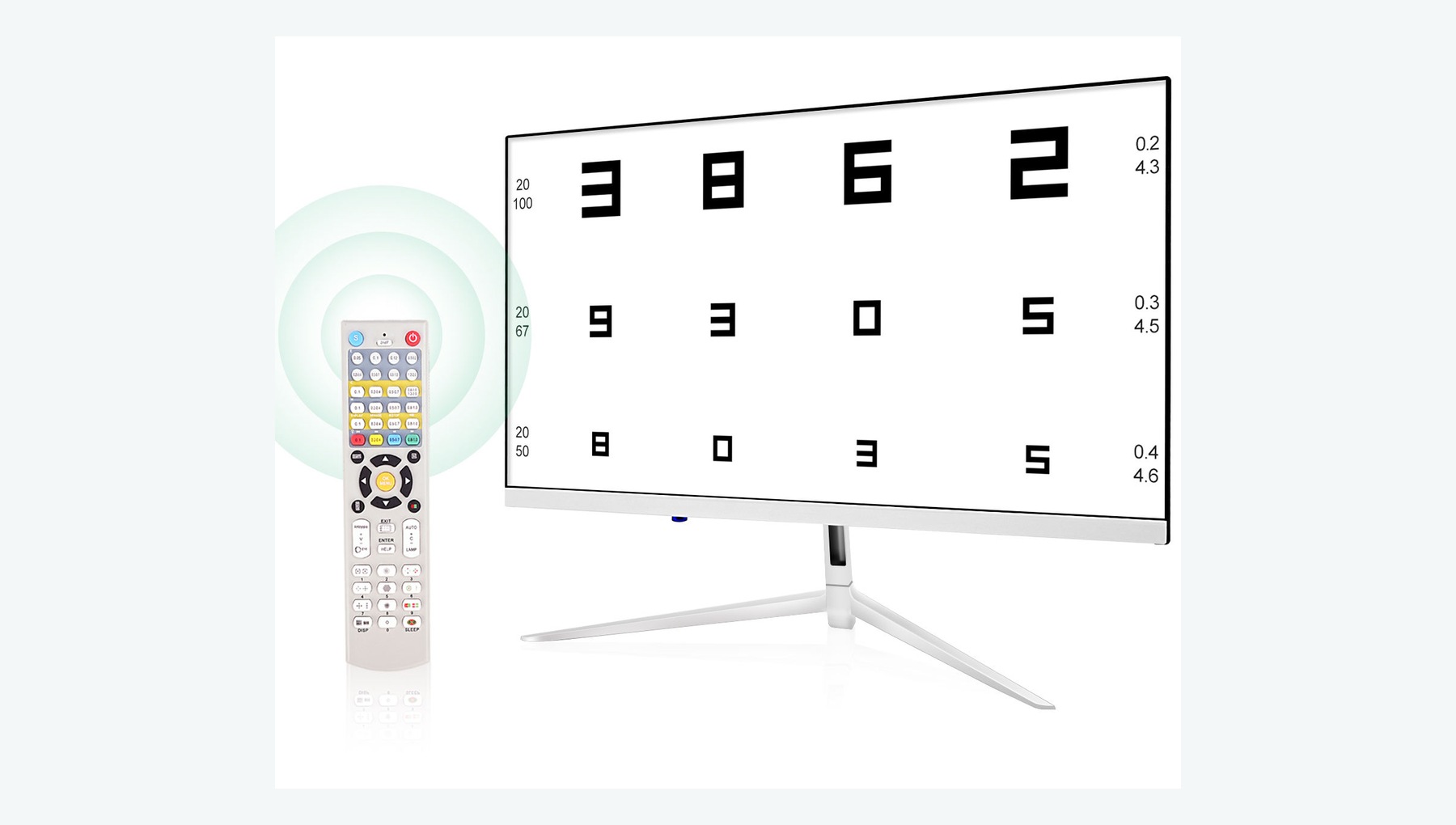
Assesses uncorrected and corrected distance and near visual acuity before and after surgery.
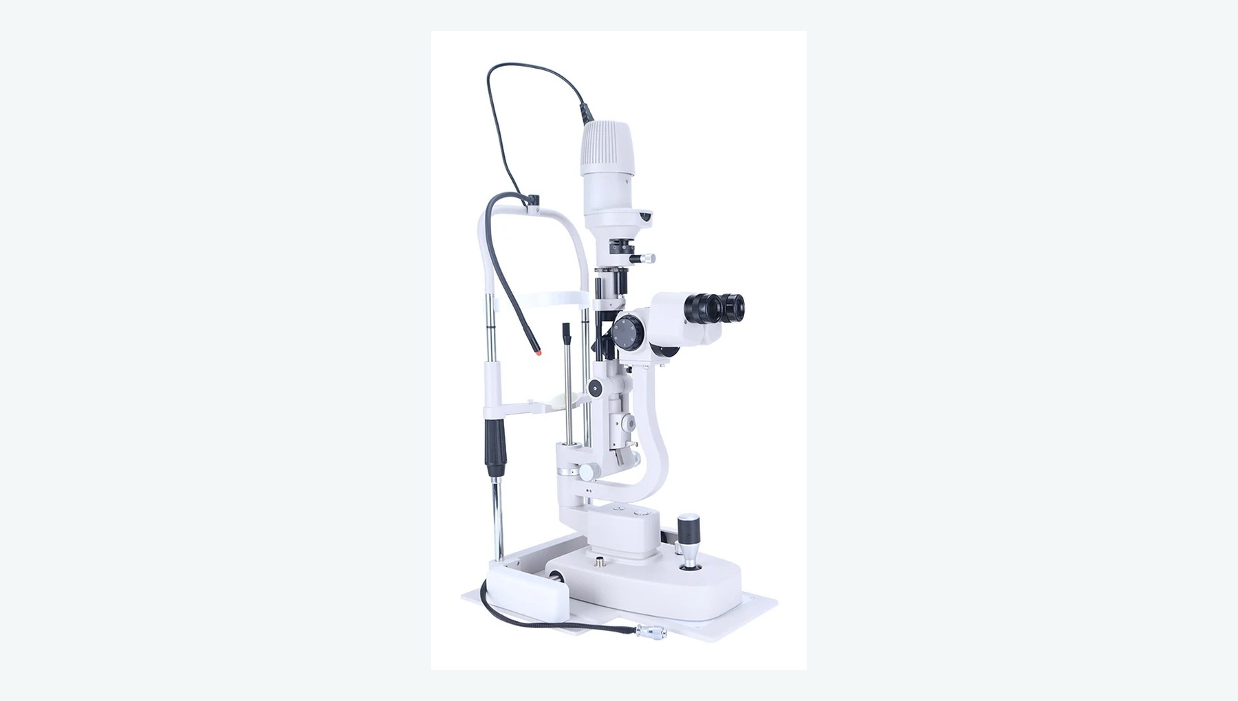
Examines the cornea, anterior chamber, pupil and lens opacity, and supports postoperative assessment of the incision and inflammation.
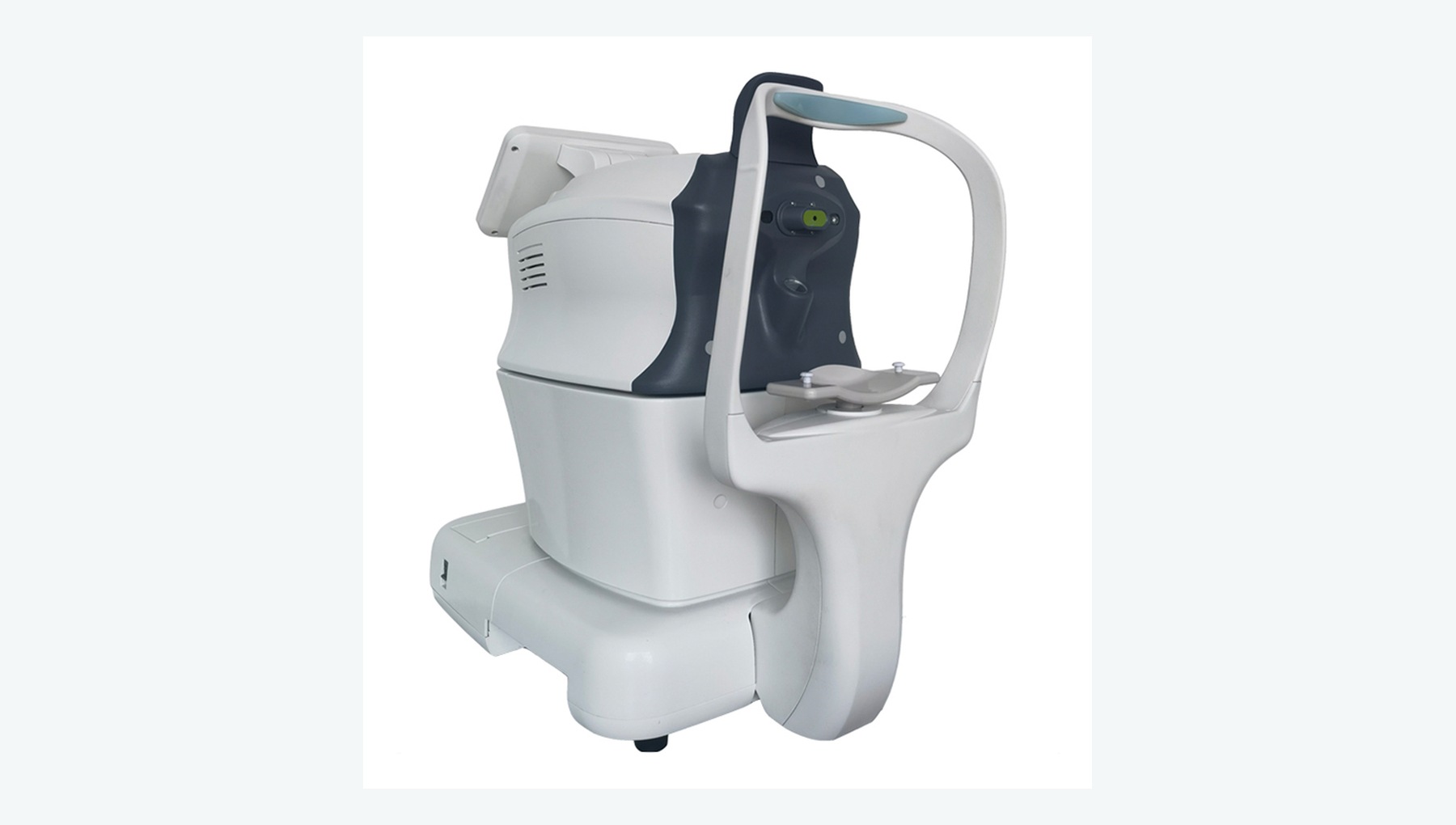
Uses an air pulse to estimate intraocular pressure rapidly without contact.
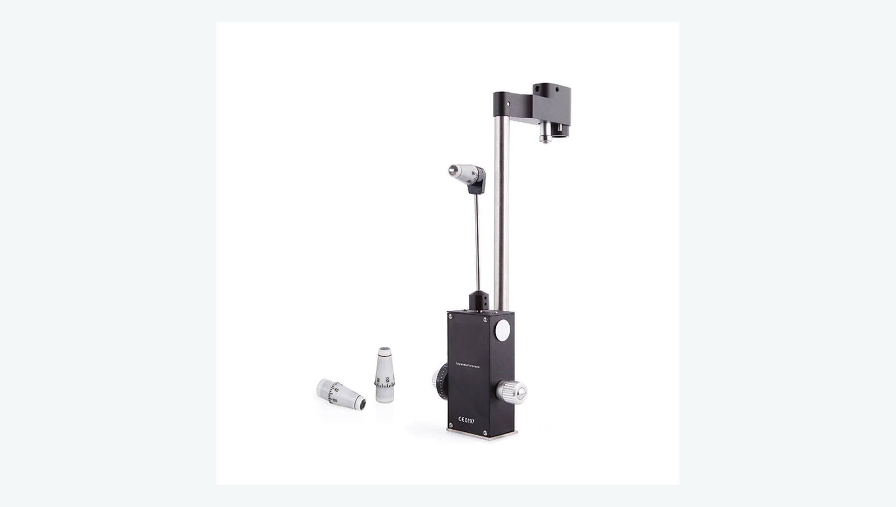
Quantifies intraocular pressure by measuring the force required to flatten a defined area of the cornea.
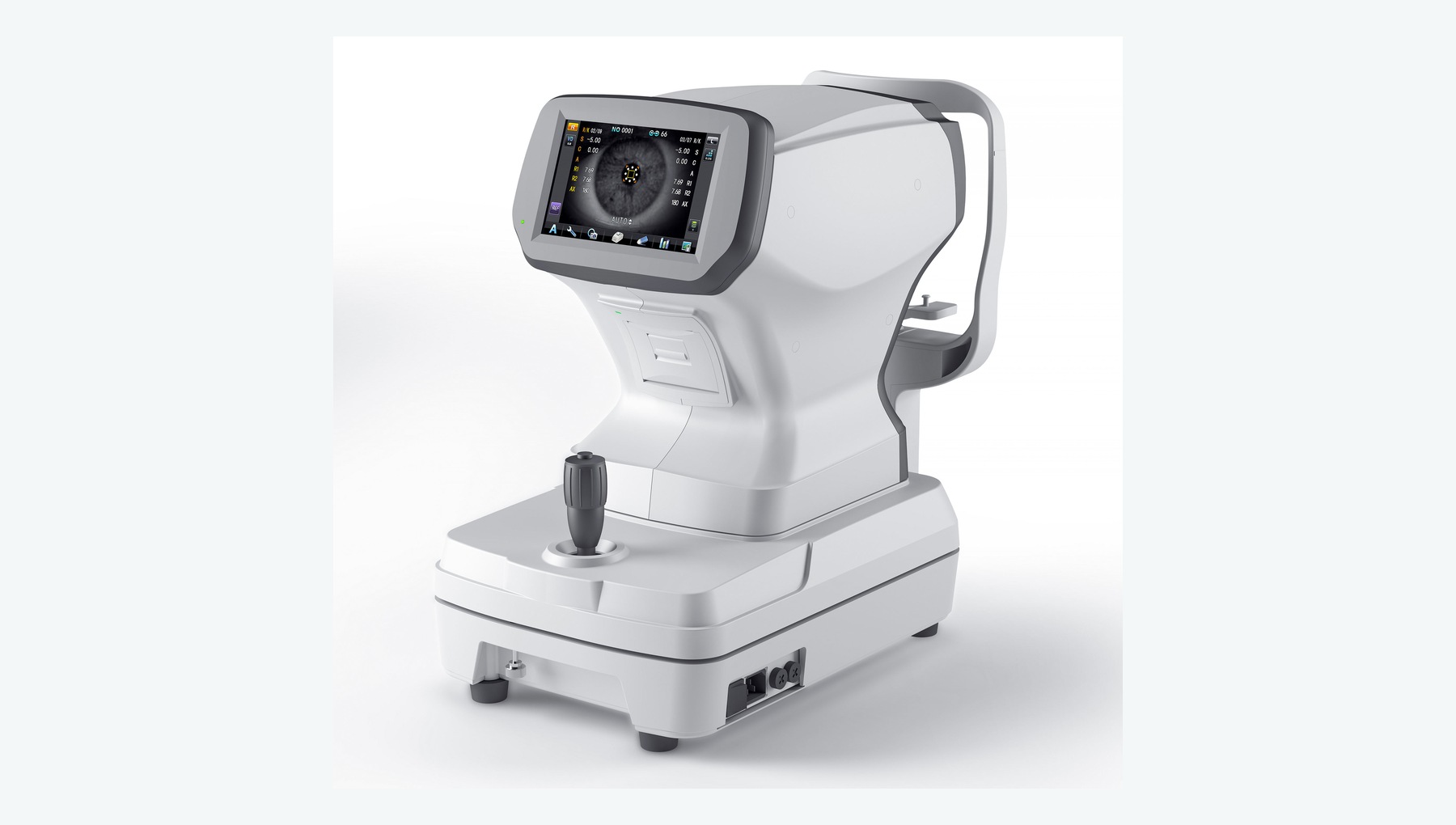
Measures objective refraction and corneal curvature to support preoperative assessment and postoperative refraction.
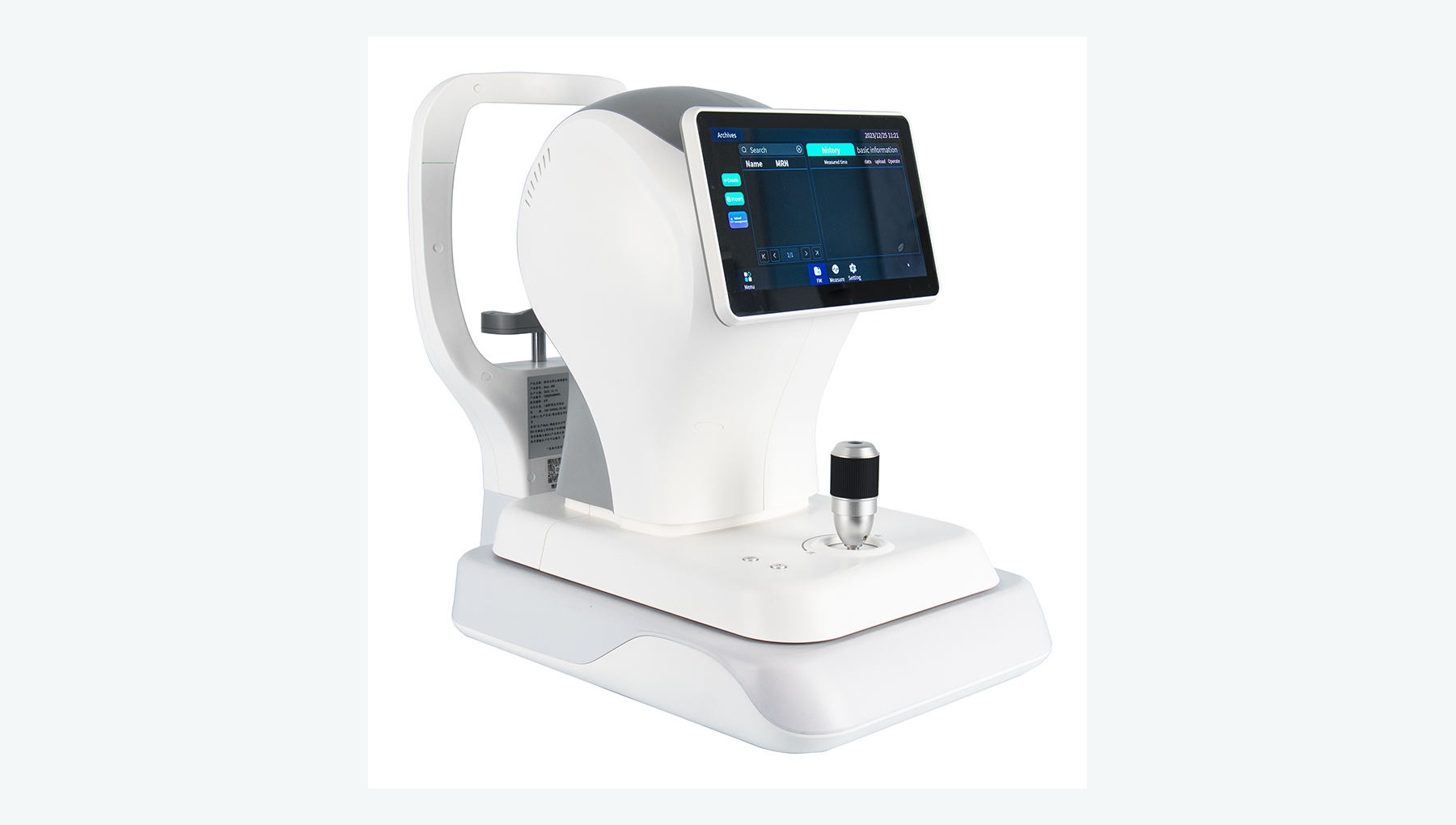
Measures axial length, corneal curvature, anterior-chamber parameters and other inputs used to calculate intraocular lens power.
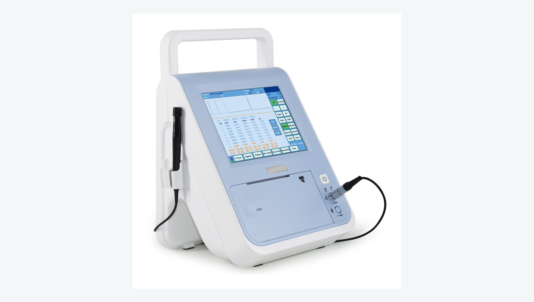
A-scan measures axial length; B-scan evaluates the vitreous and retina when opaque media prevent direct visualization.
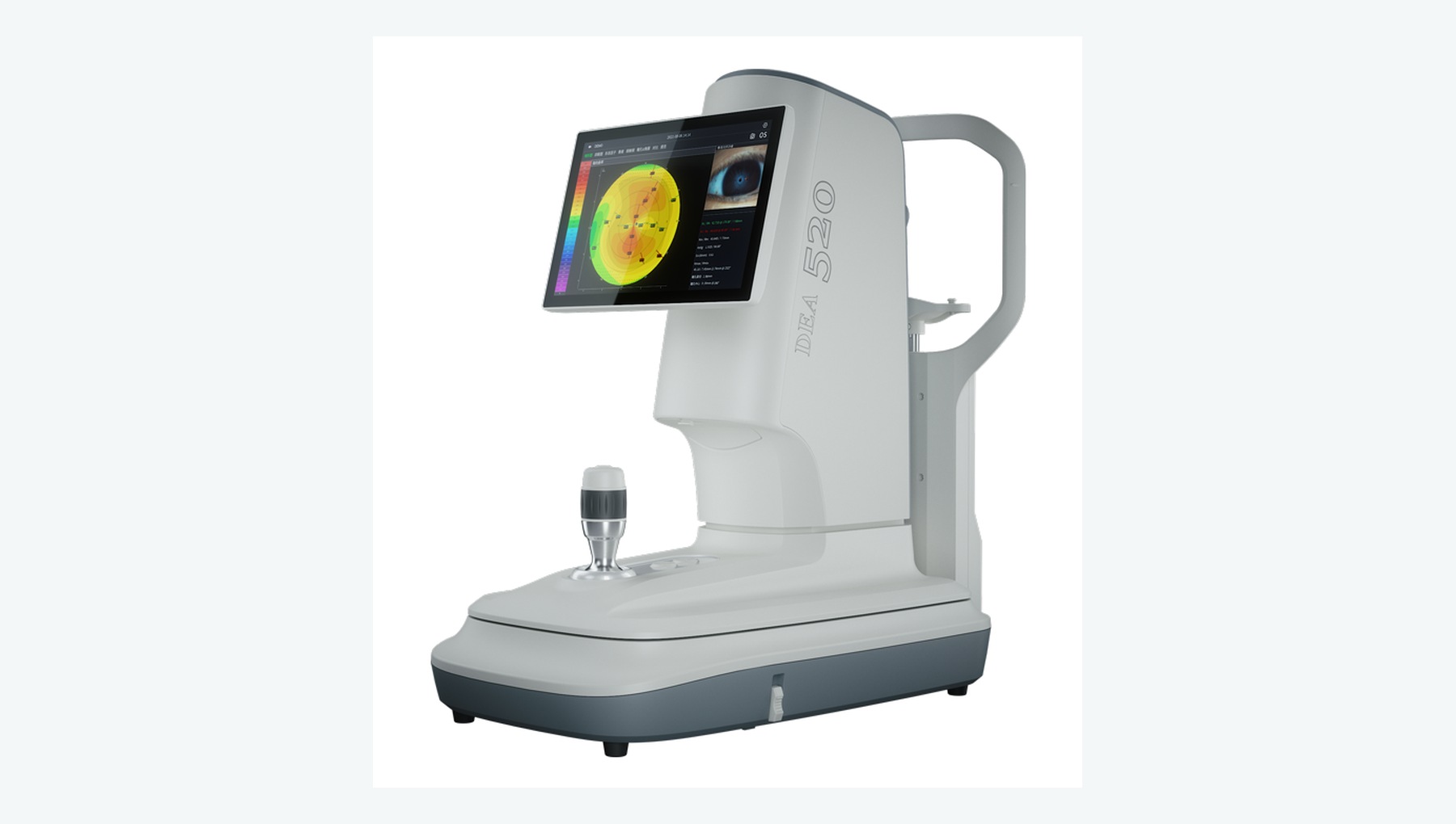
Assesses corneal curvature, regularity and astigmatism to inform more complex refractive targets.
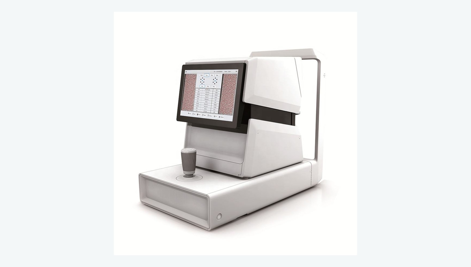
Measures corneal endothelial cell density and morphology to assess corneal risk around surgery.
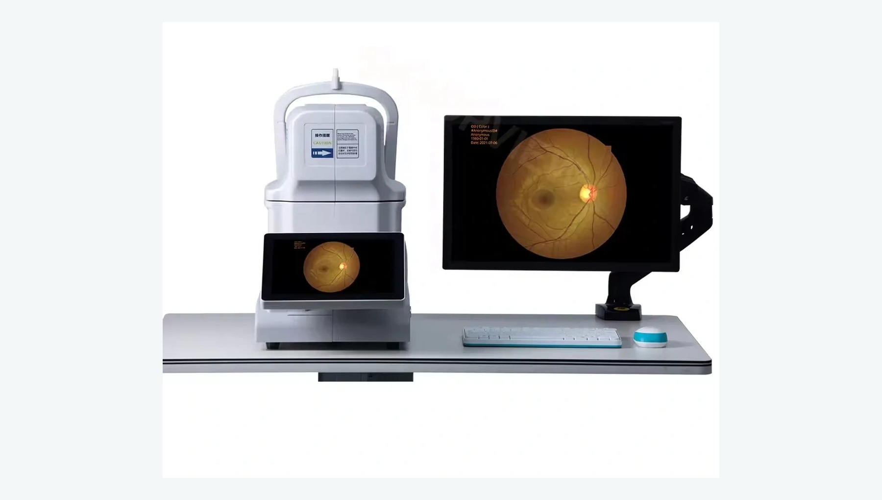
Captures color images of the optic disc, macula and retina to identify fundus disease that may limit postoperative vision.
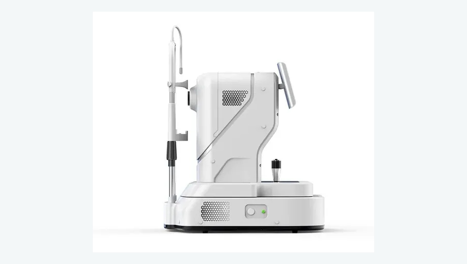
Non-invasively acquires cross-sectional, layer-by-layer images of the macula, retina and optic nerve.
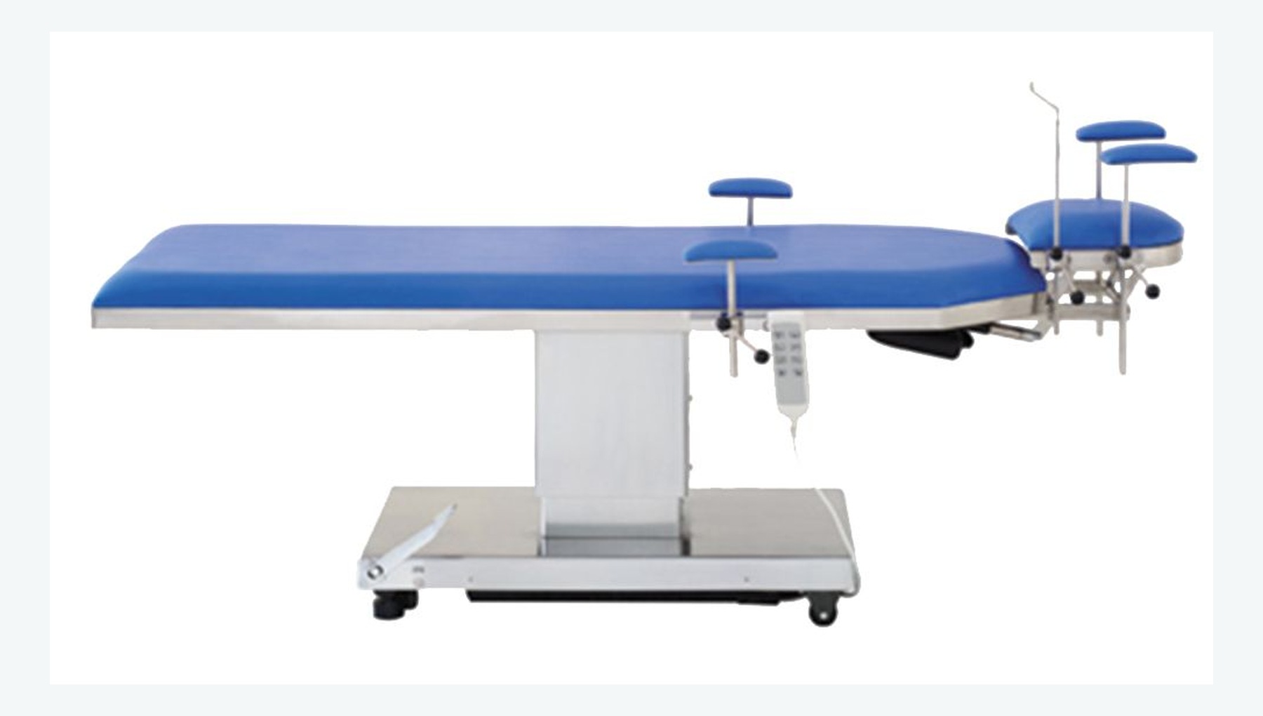
Provides stable, adjustable supine positioning and head support for microsurgery.
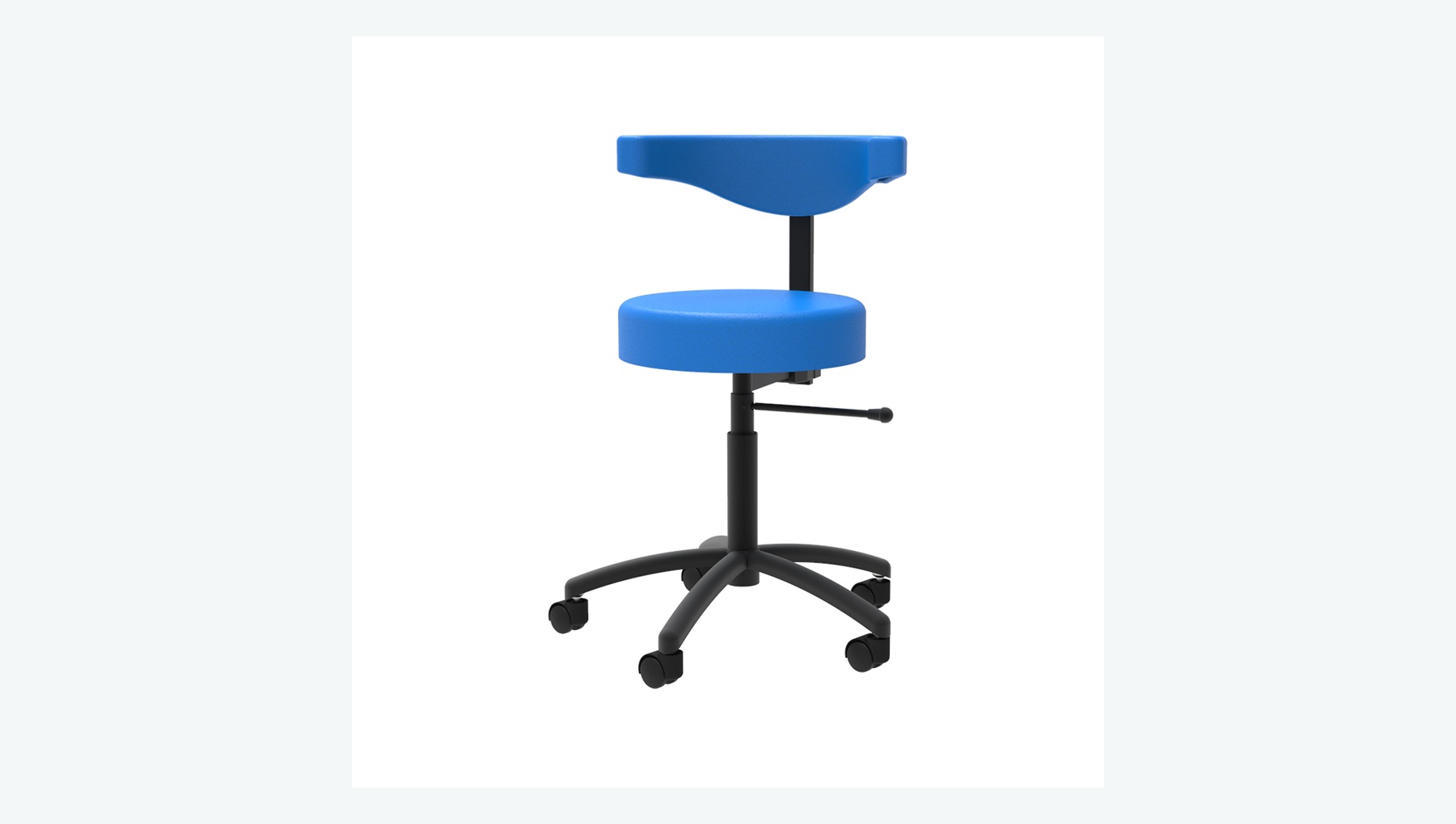
Provides adjustable height, posture and support for microsurgery, helping the surgeon maintain a stable working position.
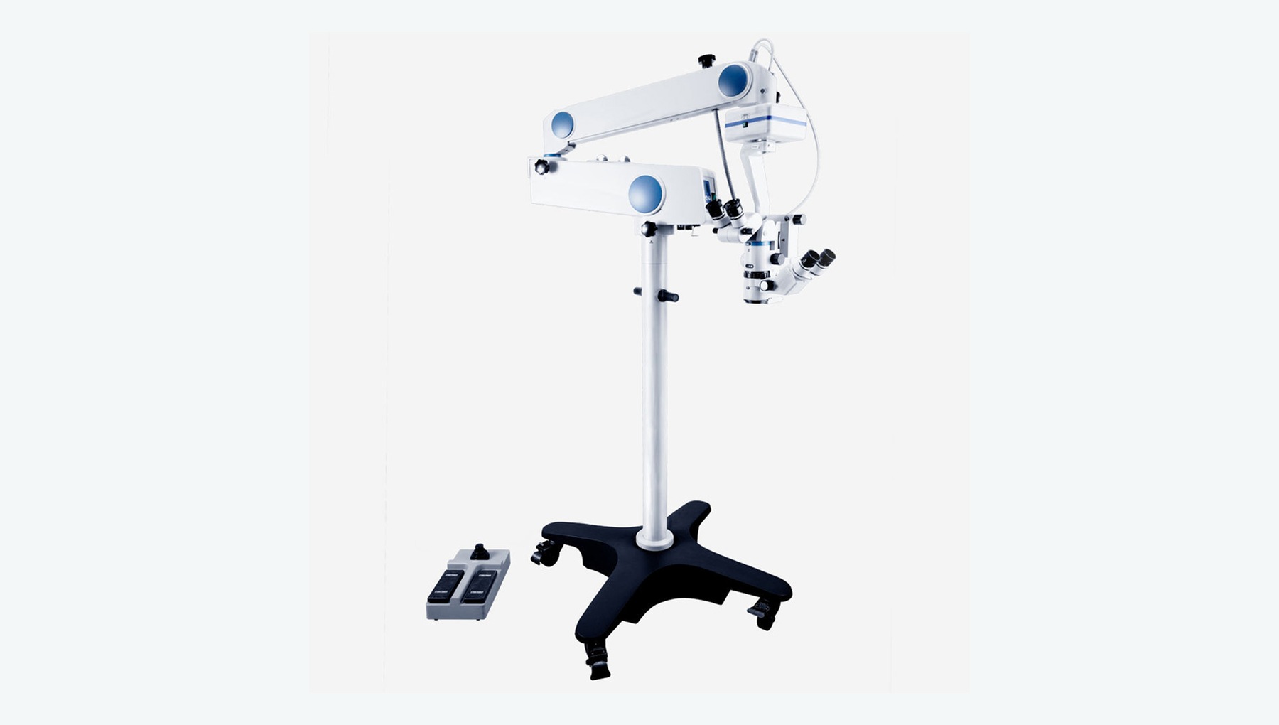
Provides coaxial illumination, stereoscopic magnification and a precise microsurgical field.
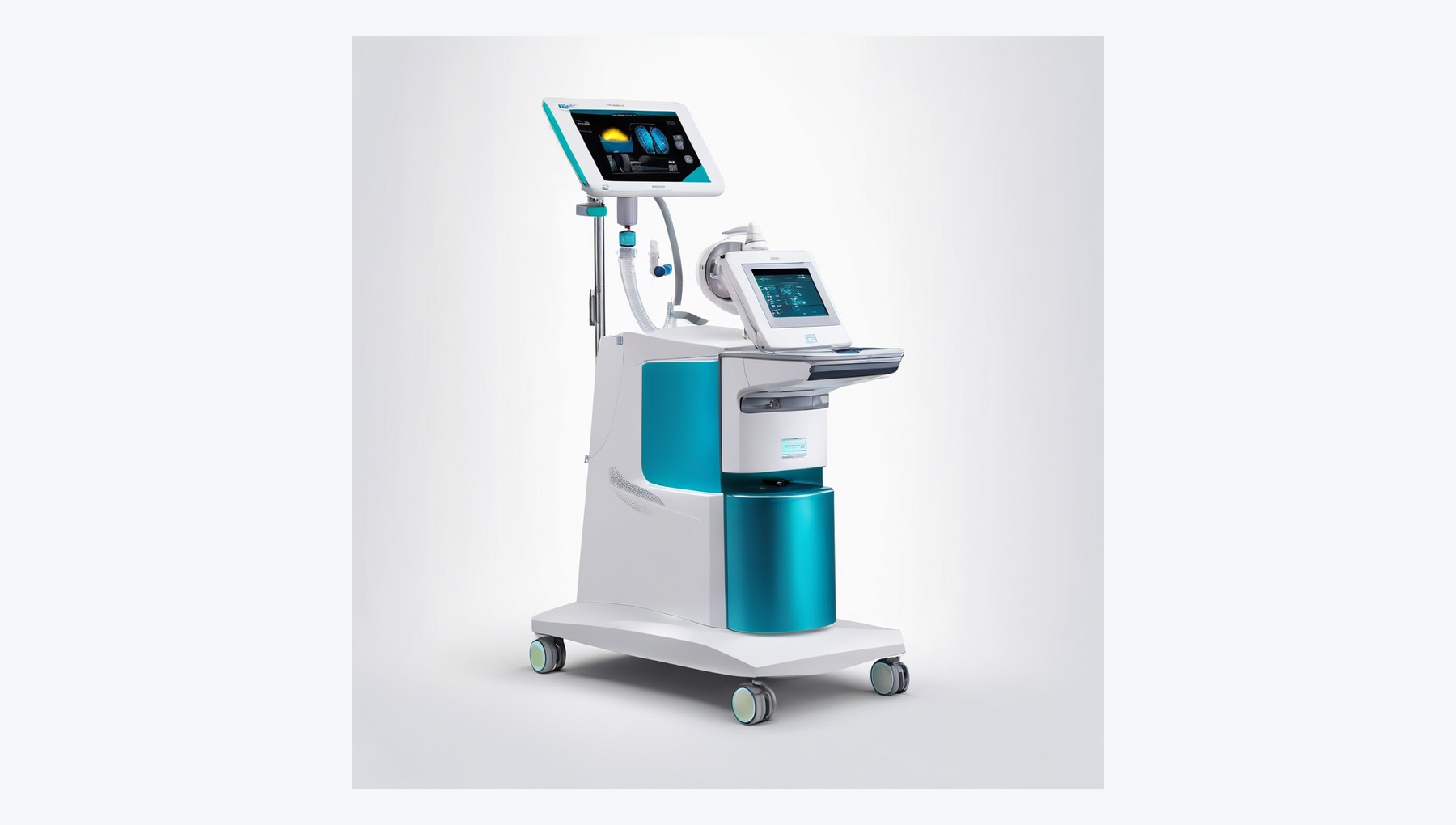
Supports ultrasonic fragmentation of the lens nucleus, irrigation and aspiration, with anterior vitrectomy capability where configured.
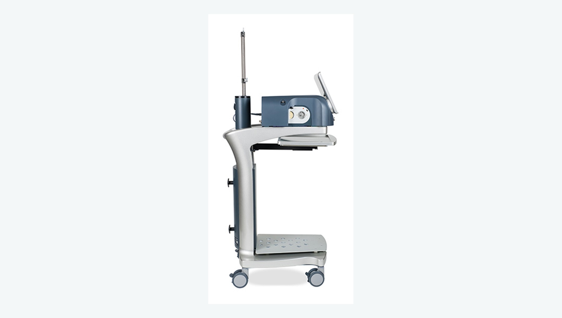
Performs anterior vitrectomy when posterior-capsule rupture, vitreous prolapse or a similar complication occurs.
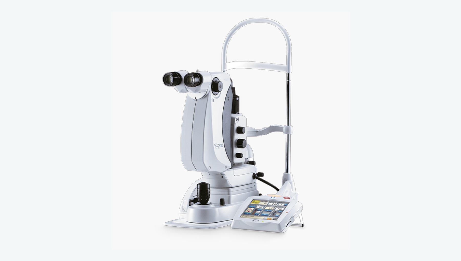
Creates a non-contact opening in the posterior capsule to restore the optical pathway.
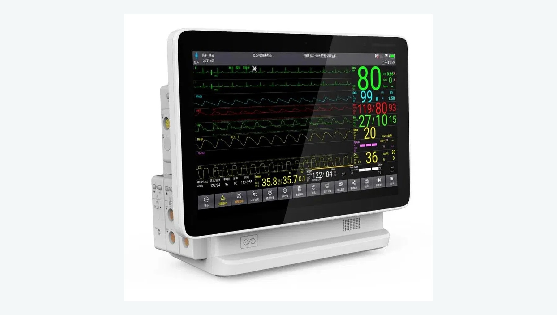
Monitors perioperative vital signs such as blood pressure, oxygen saturation and ECG.
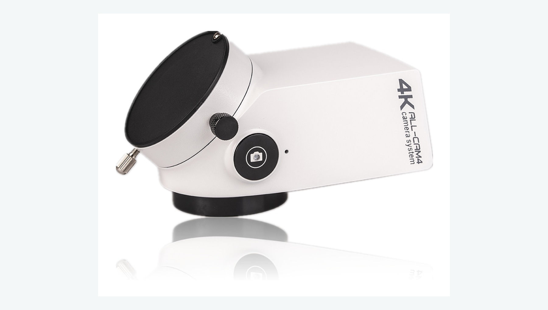
Records video from the surgical microscope for case documentation, quality review and training.
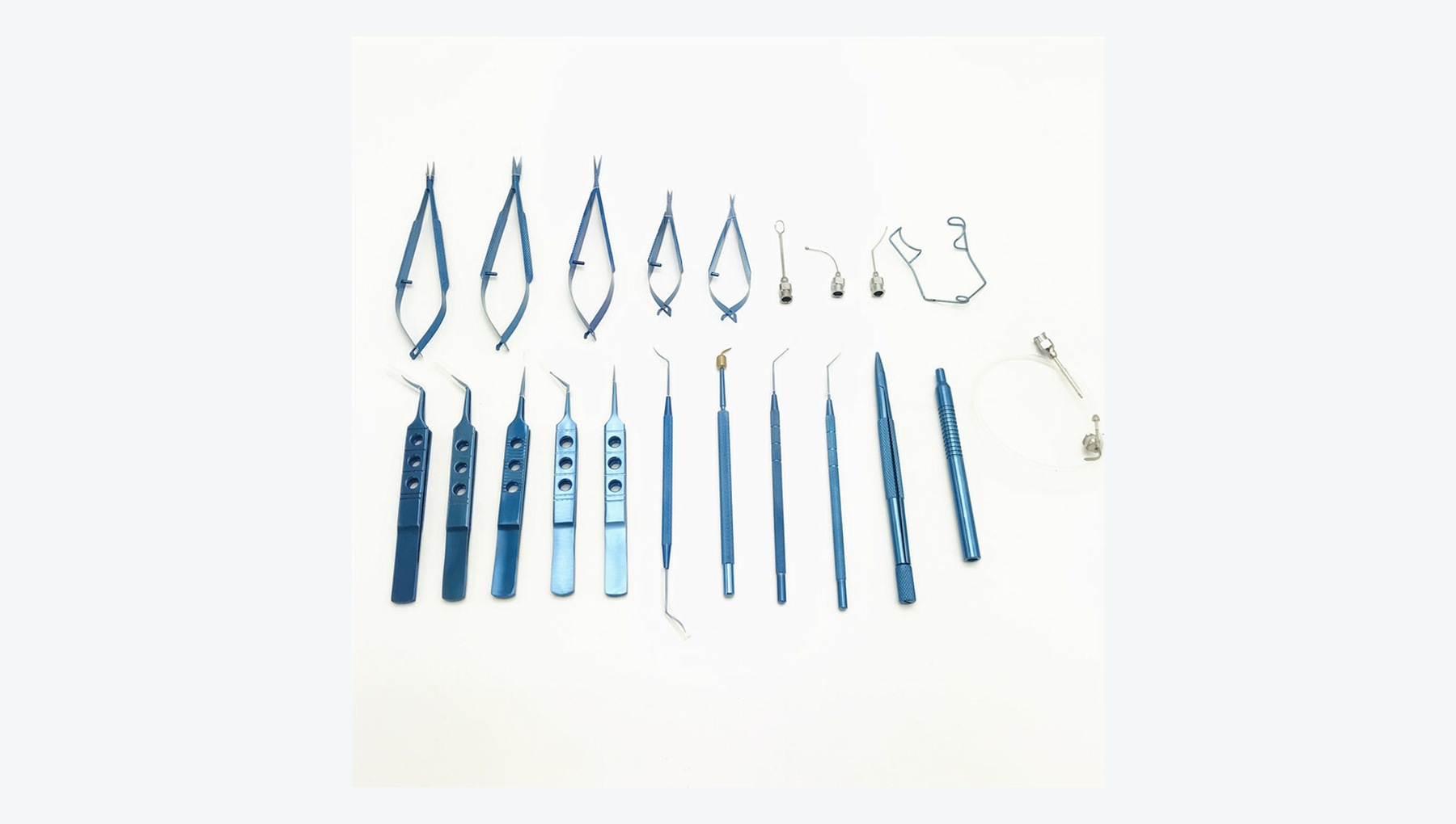
Supports lid opening, incision creation, anterior-chamber maneuvers, IOL implantation and wound management.
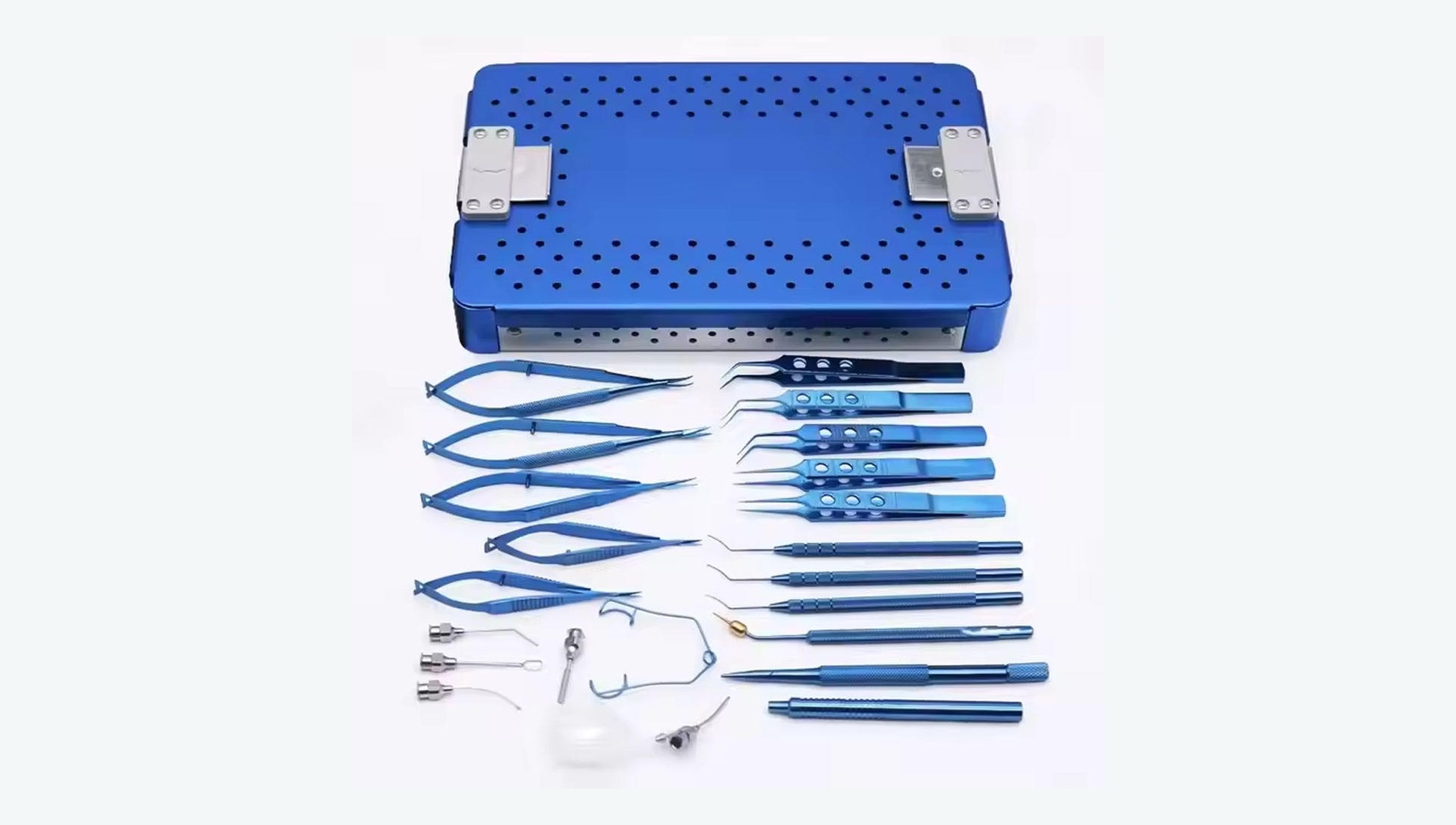
Supports clear-corneal incision, capsulorhexis, hydrodissection, nucleus disassembly, cortical cleanup and IOL implantation.
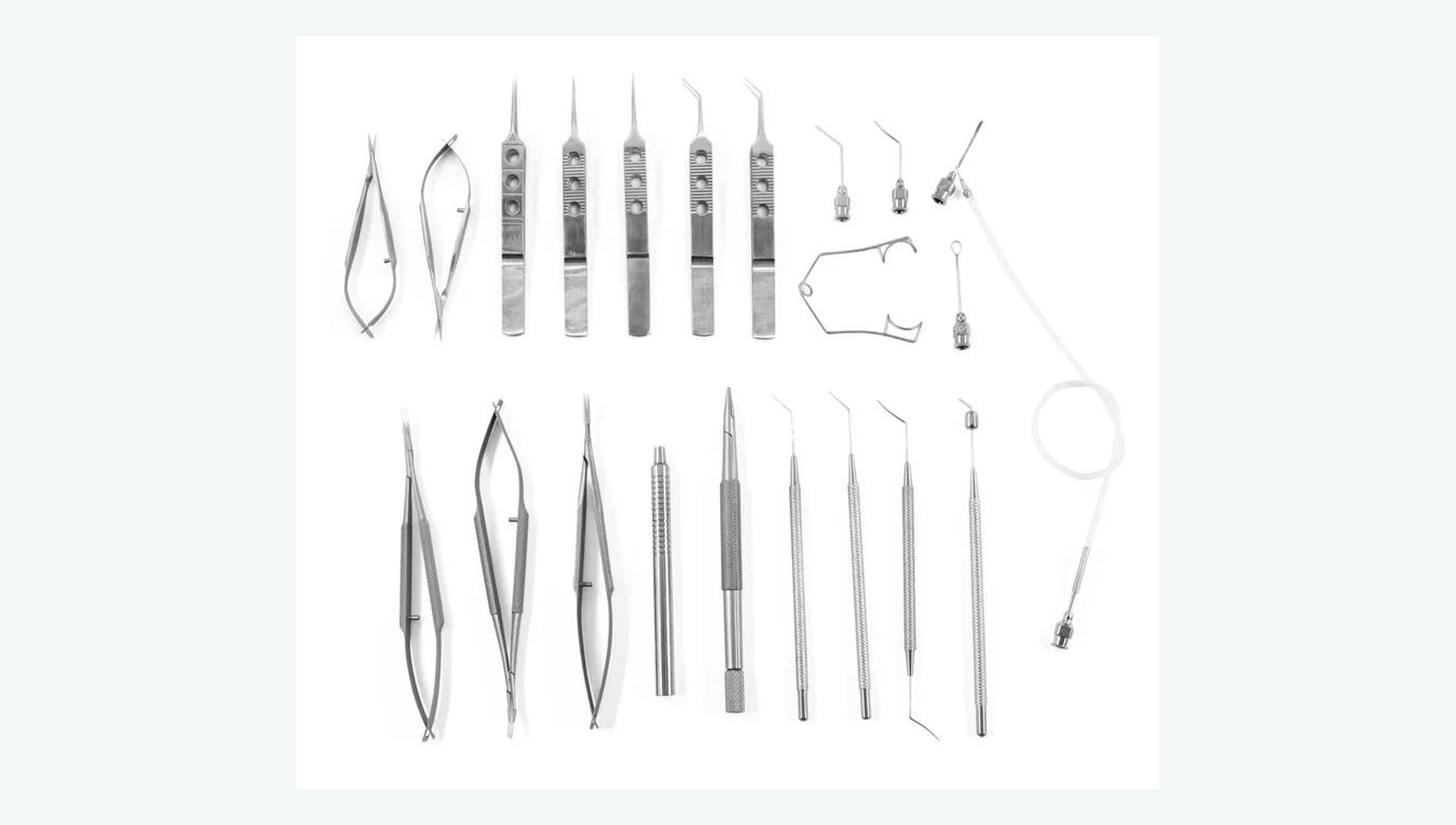
Supports creation of the scleral tunnel, nucleus delivery, cortical cleanup and IOL implantation.
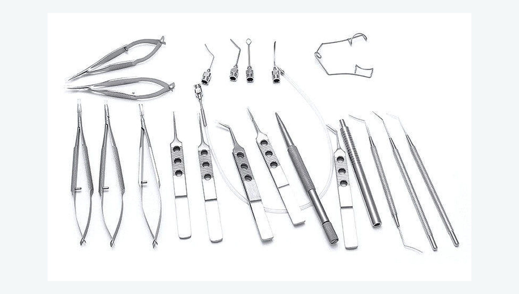
Supports large-incision extracapsular extraction, nucleus delivery, suturing and related maneuvers.
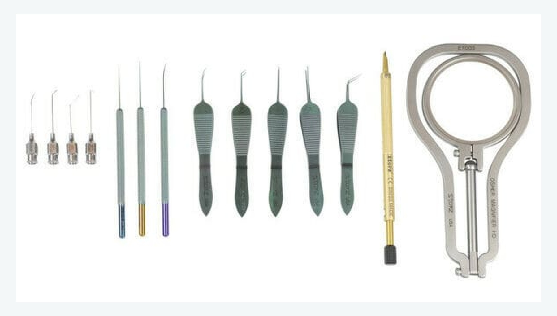
Supports small-pupil expansion, capsular-bag stabilization and cases involving iris or zonular abnormalities.
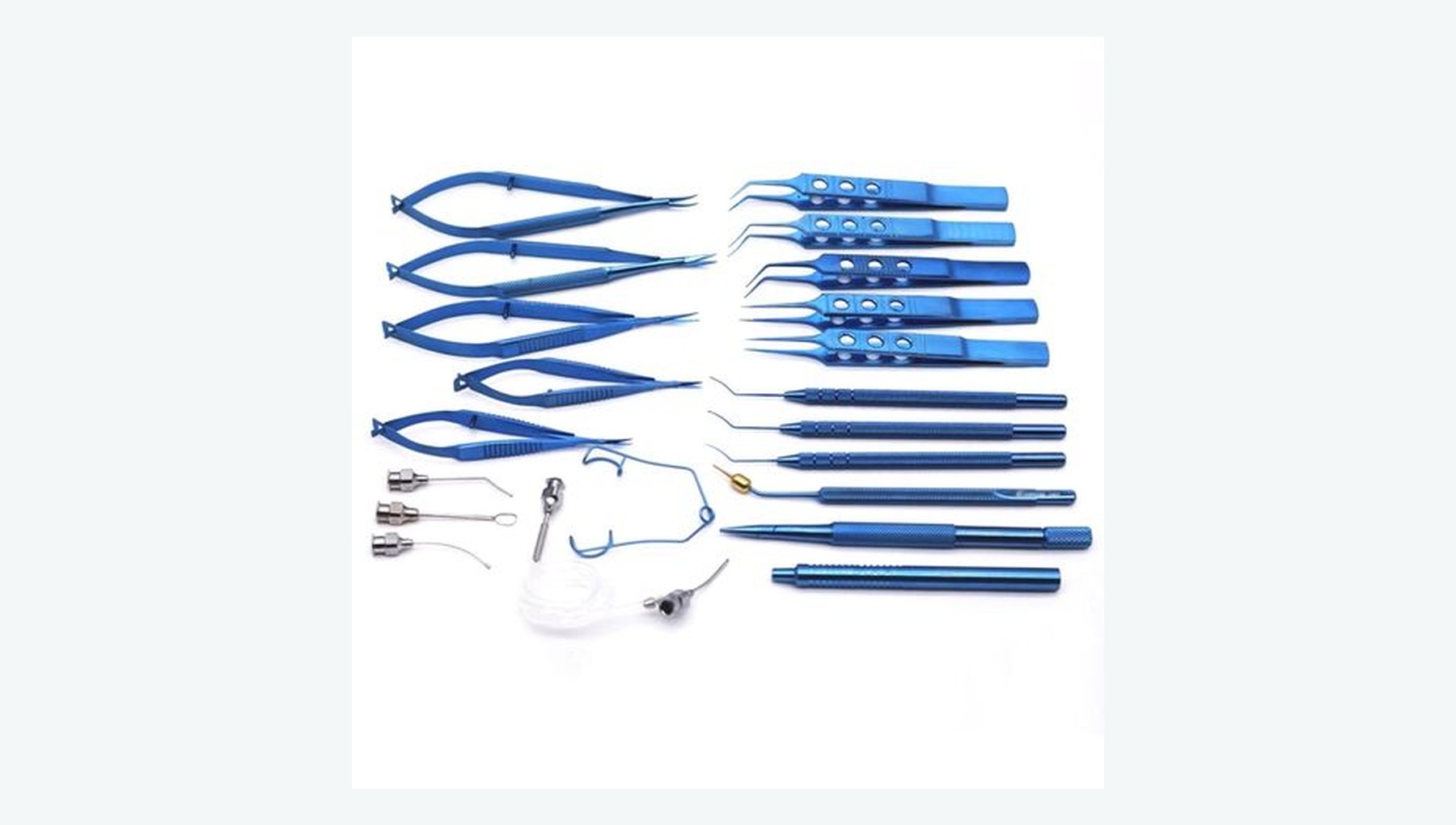
Supports management of posterior-capsule rupture, vitreous prolapse and vitreous in the anterior segment.
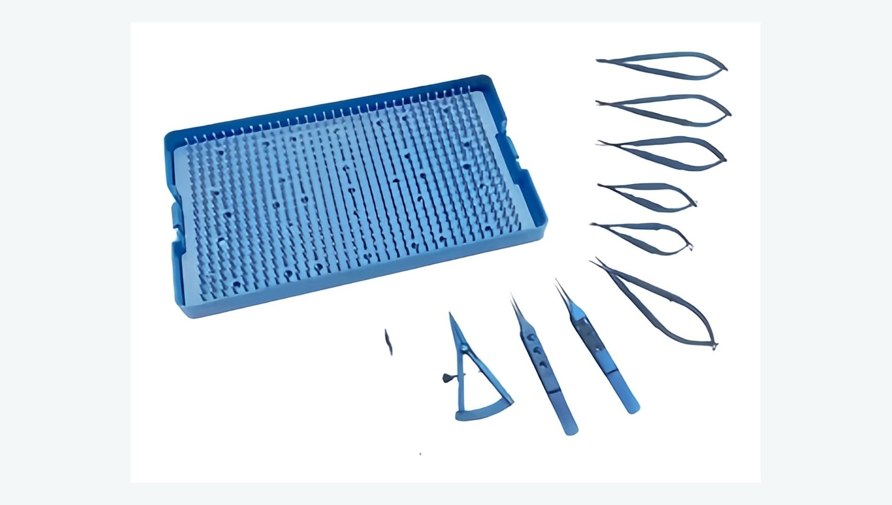
Supports foldable IOL insertion, positioning and, when required, management or removal of the implant.
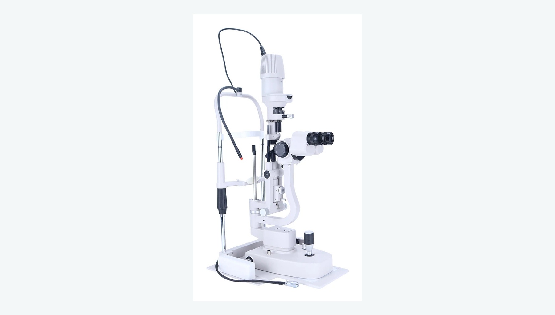
Provides magnified anterior-segment examination and, with a non-contact fundus lens, assessment of the vitreous, macula and optic nerve.
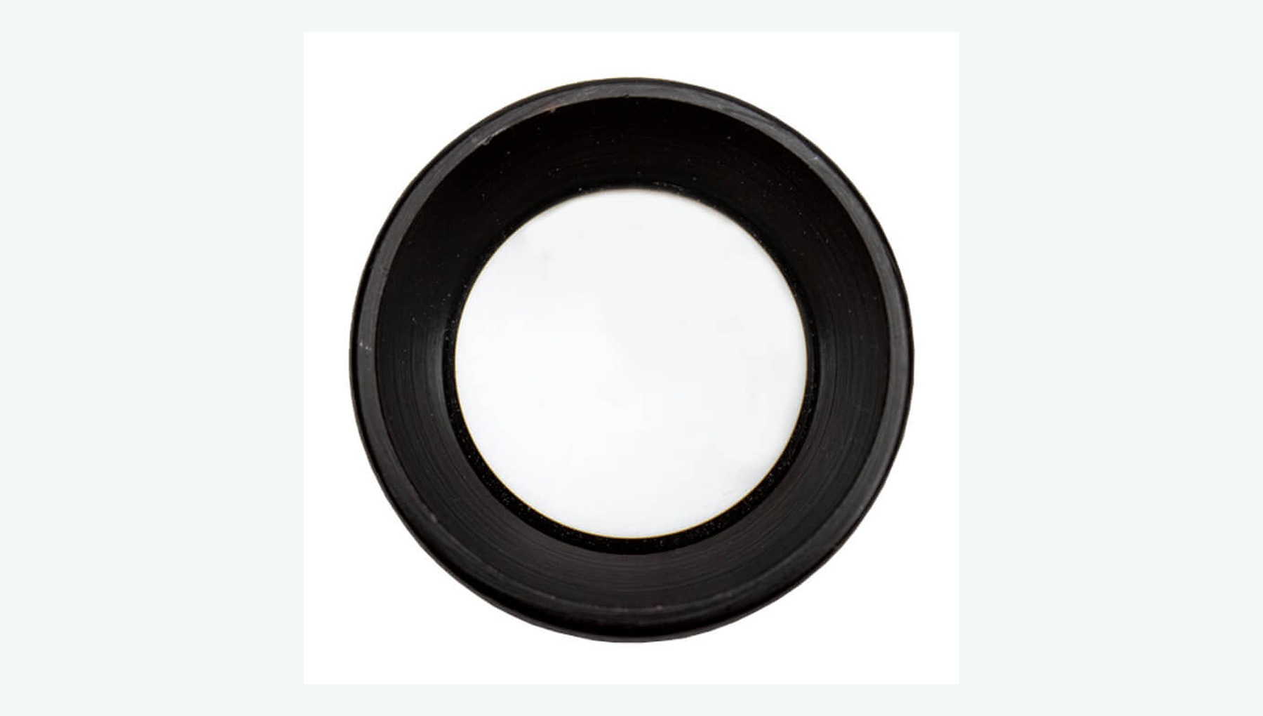
Used with a slit lamp to create a stereoscopic fundus view of the macula, optic disc and posterior retina.
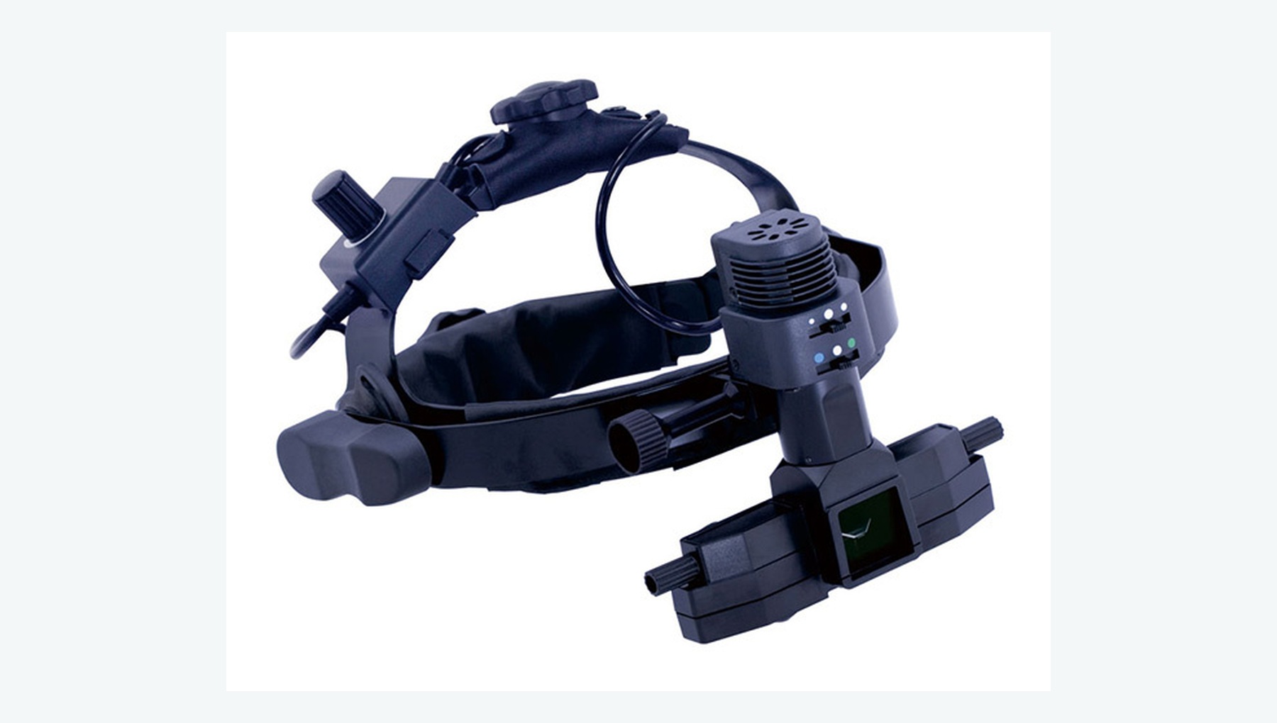
Provides a wide, dynamic fundus examination and can be combined with scleral depression to inspect the peripheral retina.
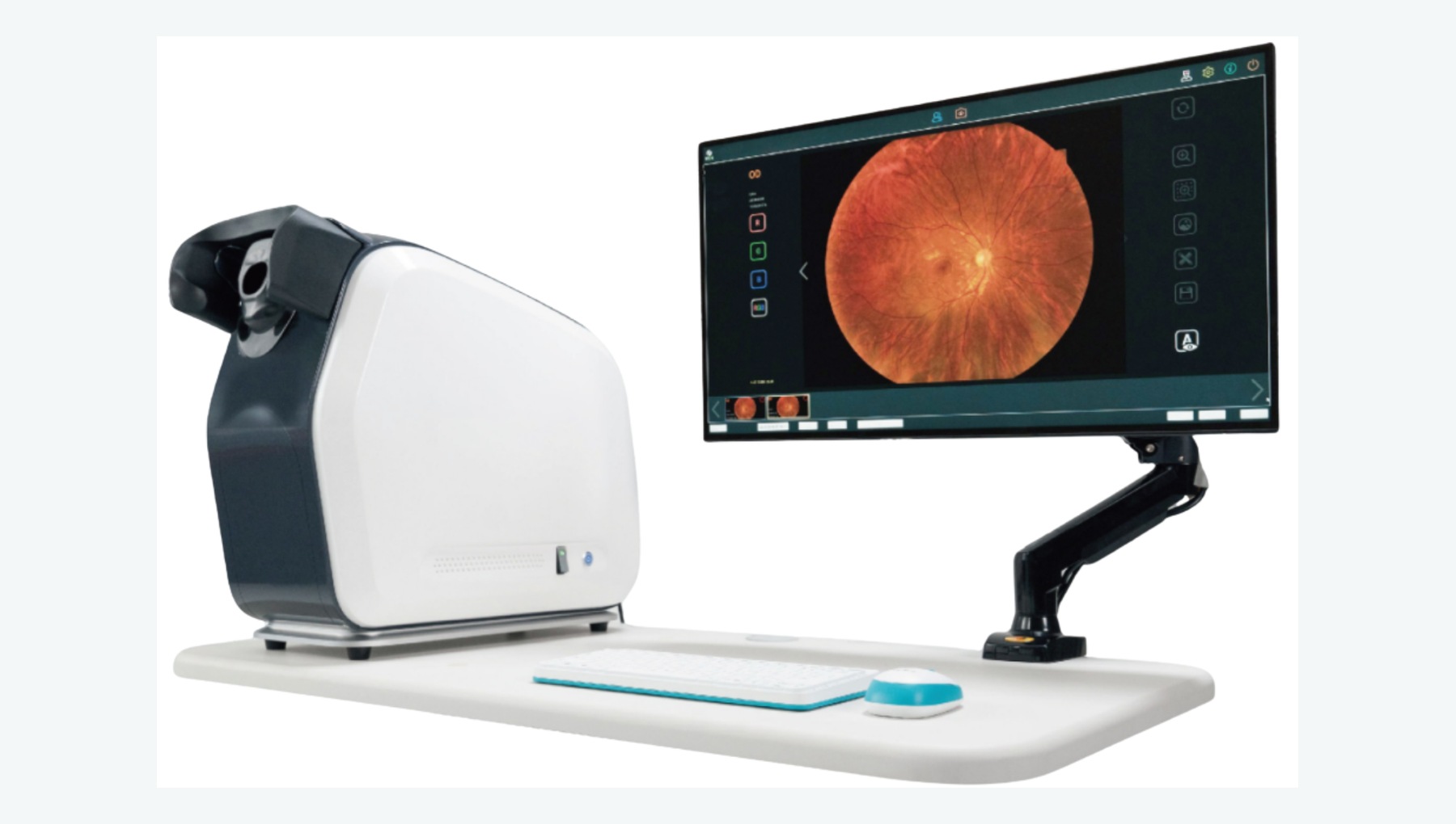
Captures a broad view of the retina in a single image or montage, with emphasis on peripheral lesions.
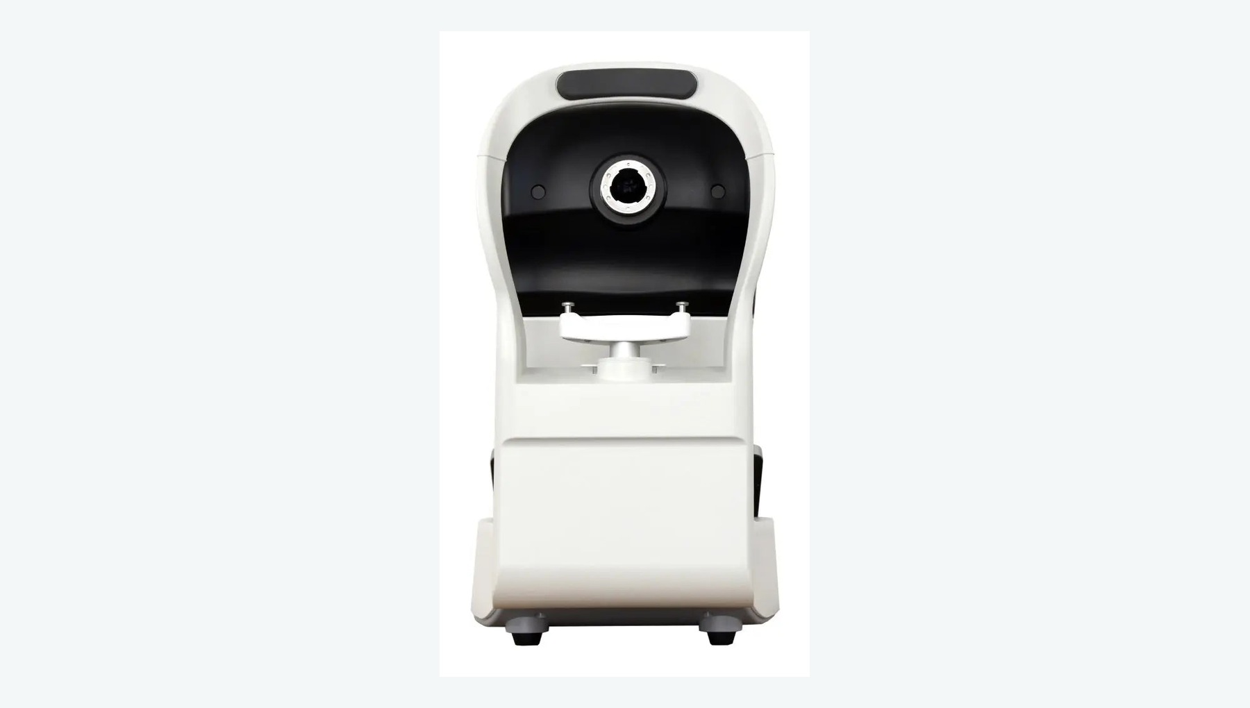
Captures images of the optic disc, macula and retina for screening, documentation and longitudinal comparison.
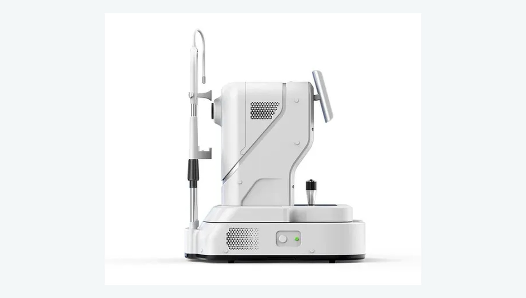
Non-invasively acquires layered cross-sectional images of the macula and retina to visualize edema, holes and epiretinal membranes.
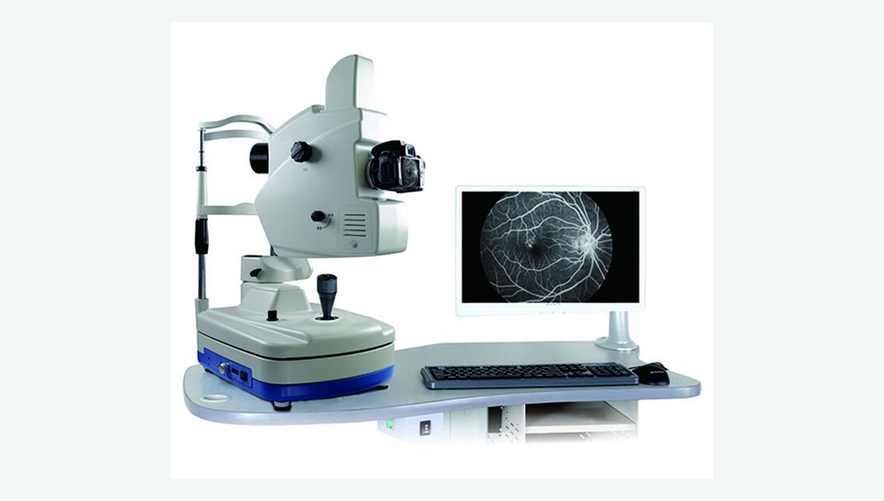
Performs fluorescein angiography and, where supported by the system configuration, indocyanine green angiography.
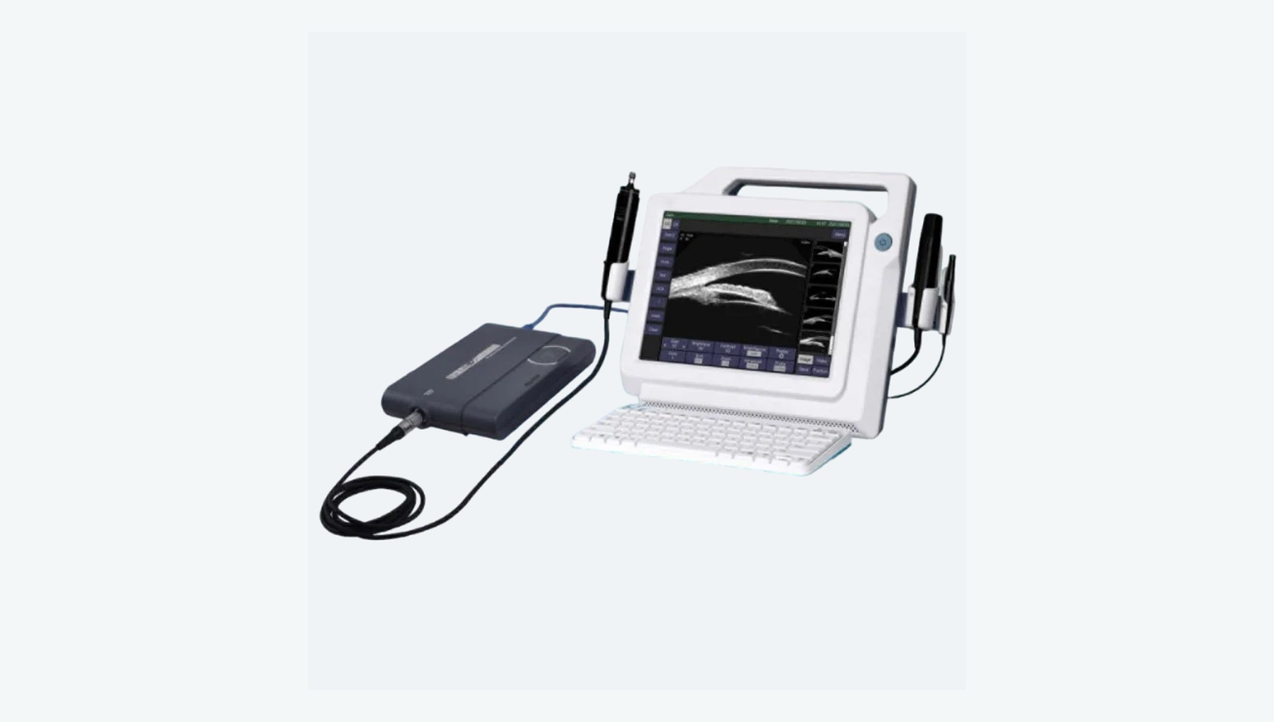
Assesses the vitreous, retina and intraocular masses when media opacity prevents visualization of the fundus.
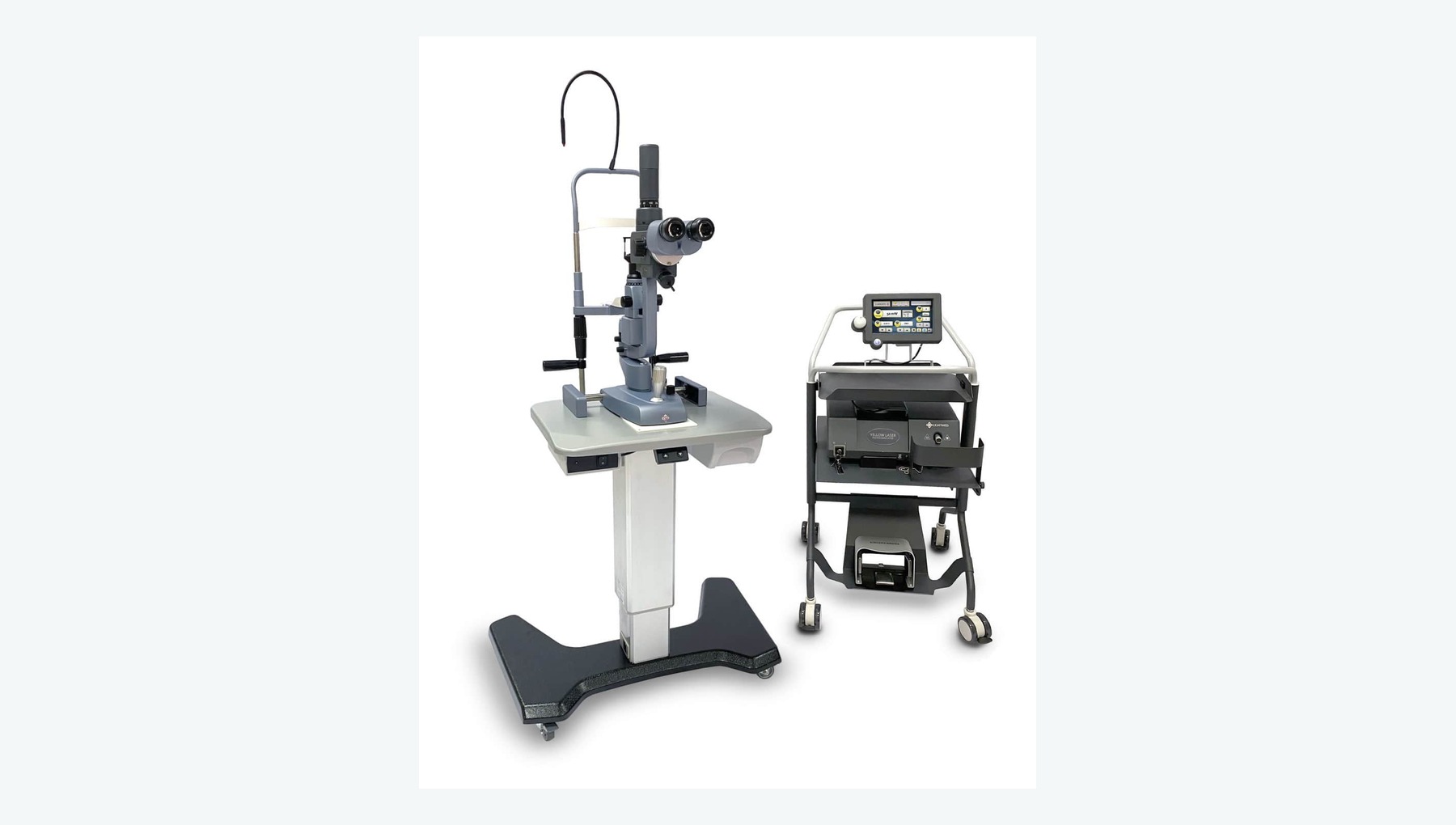
Delivers retinal laser photocoagulation through a slit-lamp or indirect-ophthalmoscope delivery system.
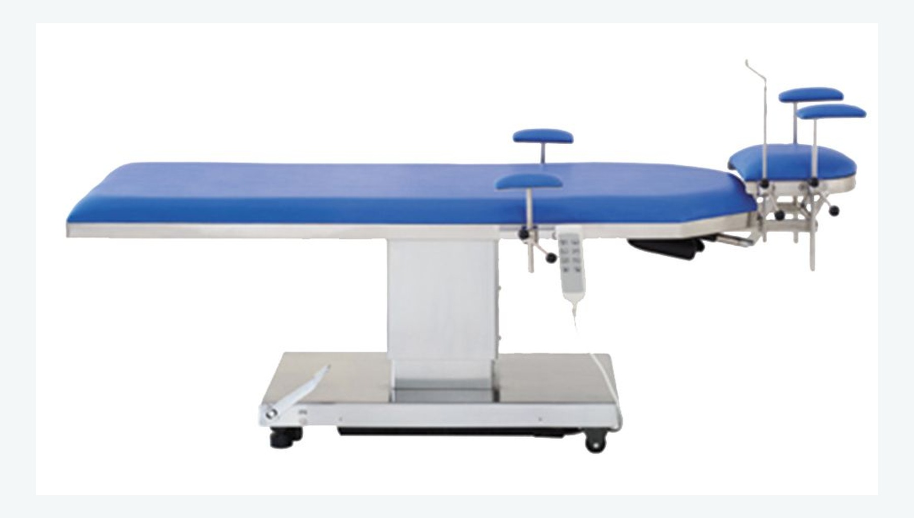
Provides stable, adjustable supine positioning and head support for microsurgery.
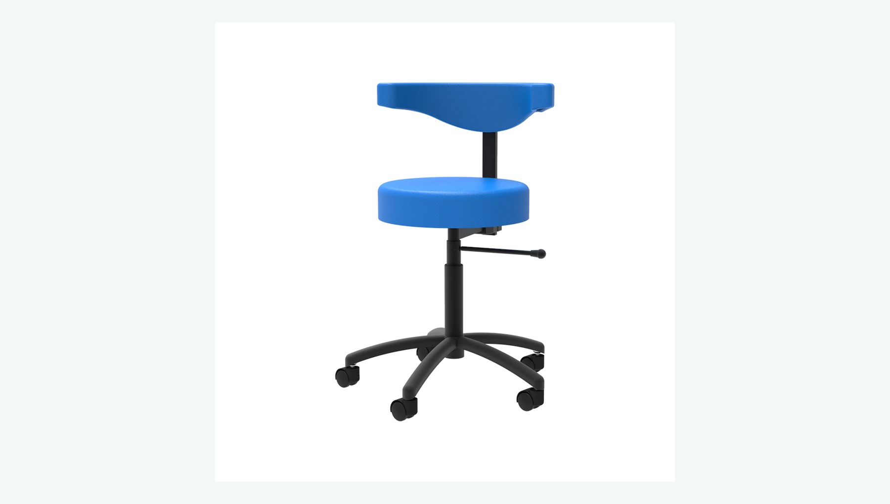
Provides adjustable height, posture and support for microsurgery, helping the surgeon maintain a stable working position.
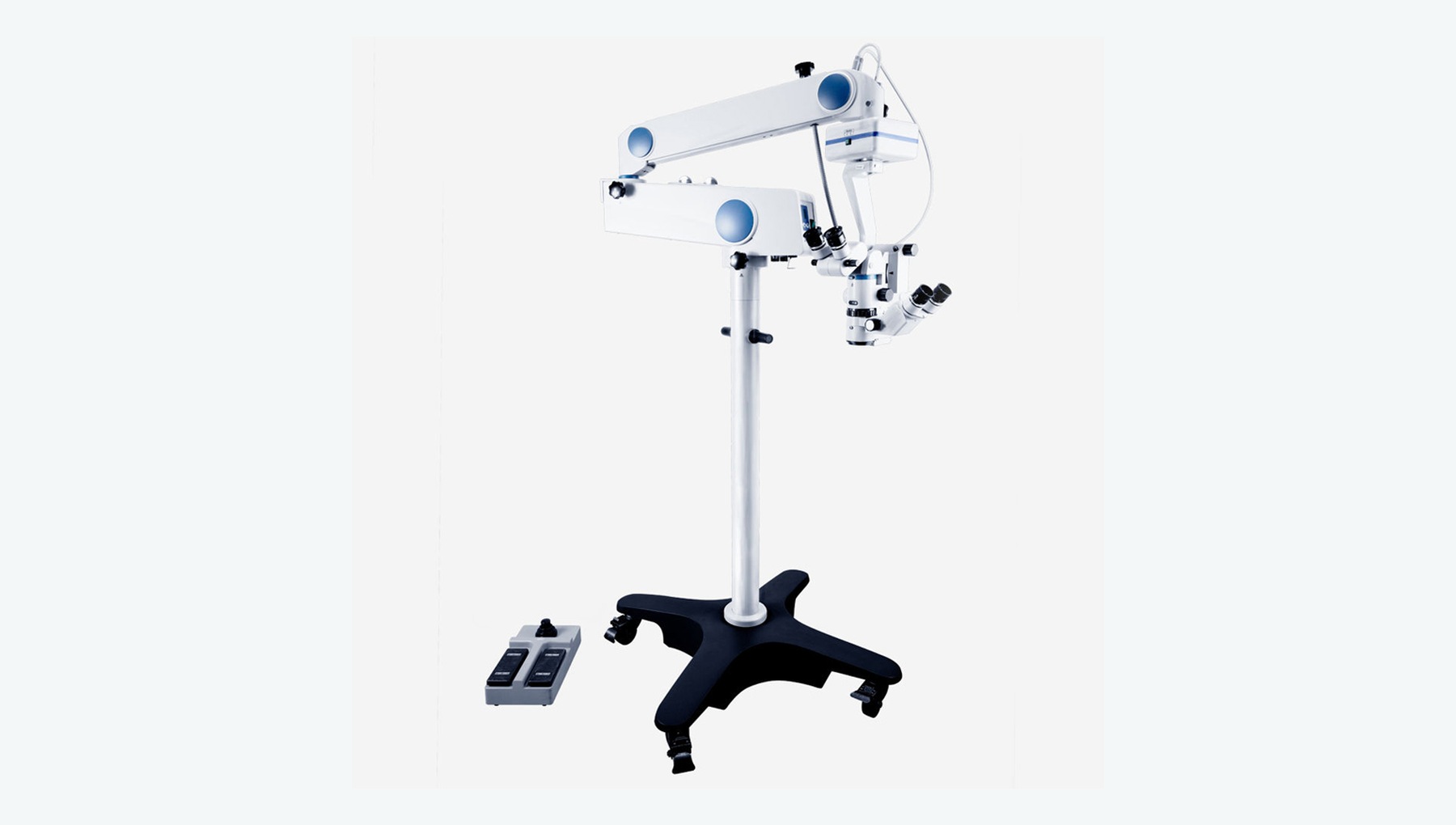
Provides high-resolution stereoscopic magnification, coaxial illumination and a clear surgical field.
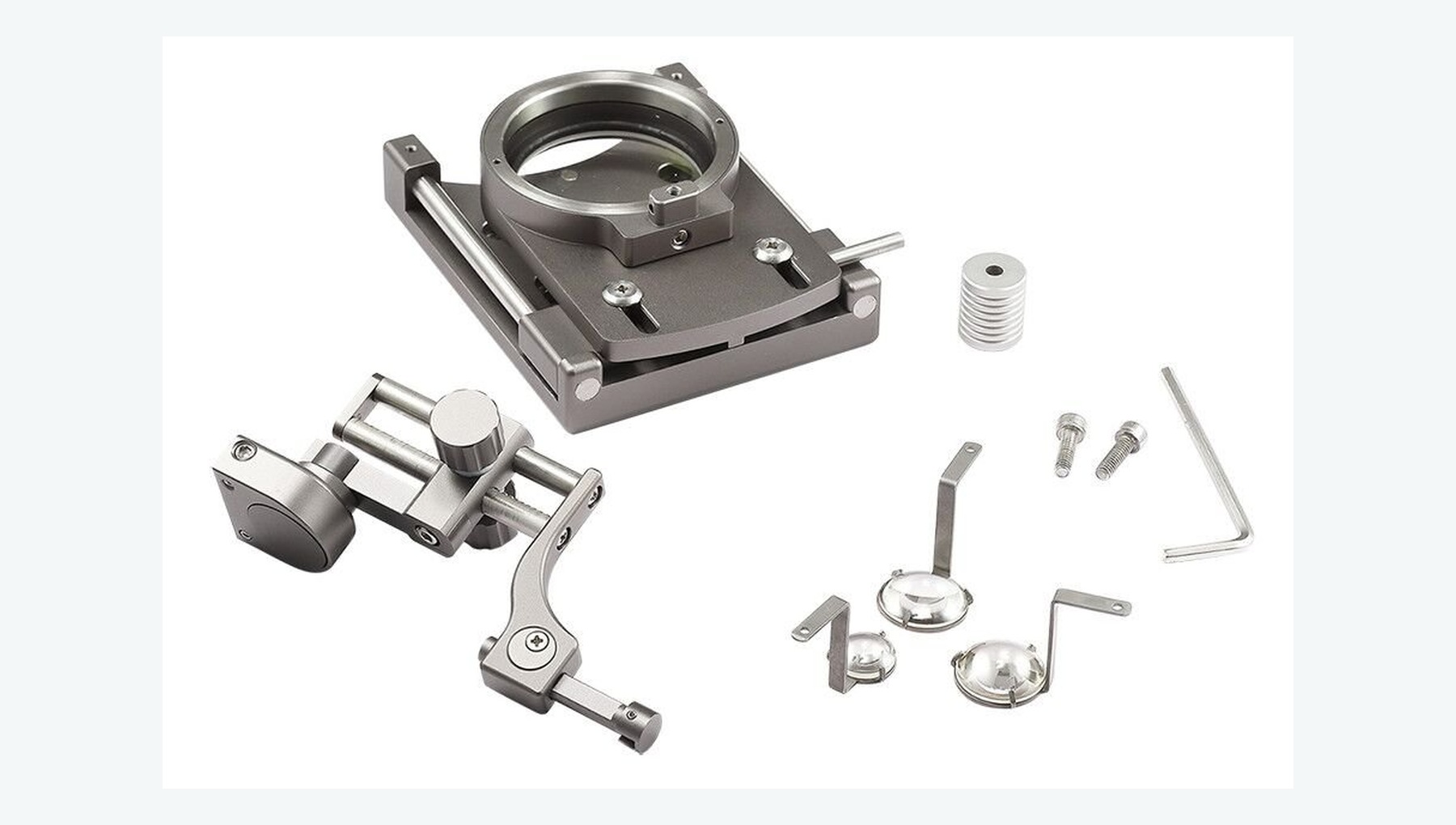
Provides a wide retinal view from the periphery to the posterior pole under the operating microscope.
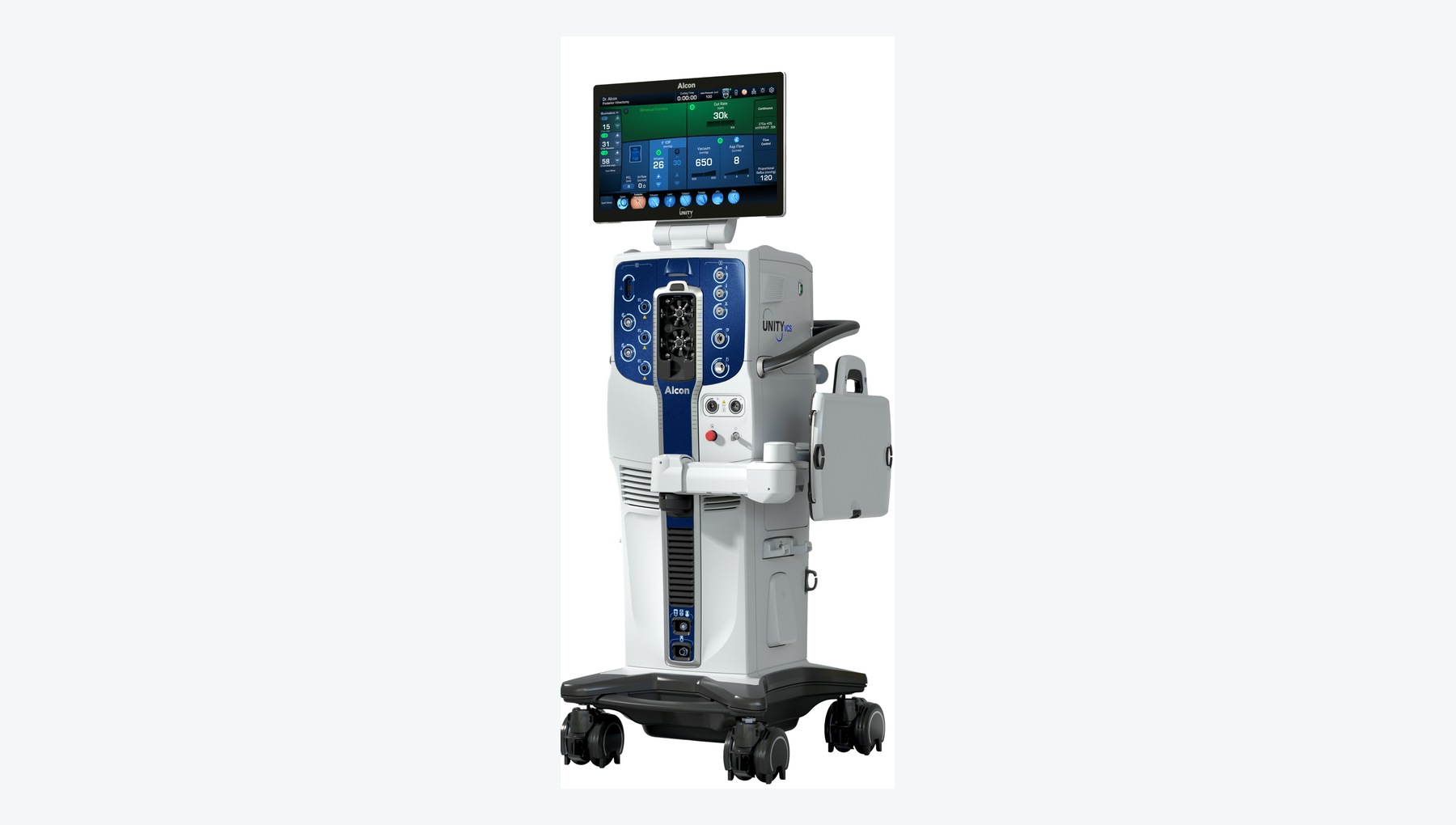
Provides infusion, vitreous cutting, aspiration, fluid-air exchange and intraocular pressure control.
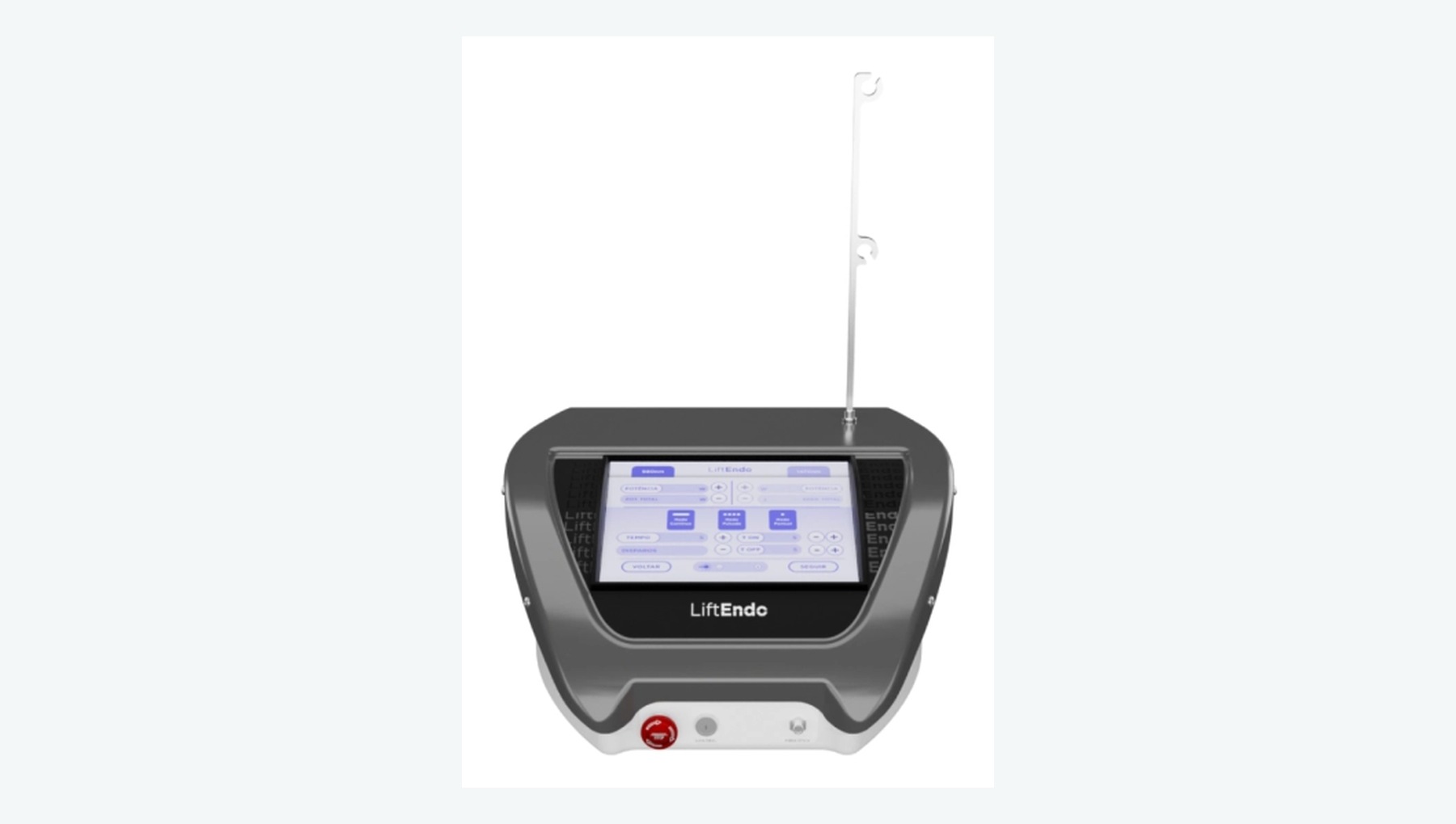
Applies intraocular laser during vitrectomy to treat retinal tears and perform retinal photocoagulation.
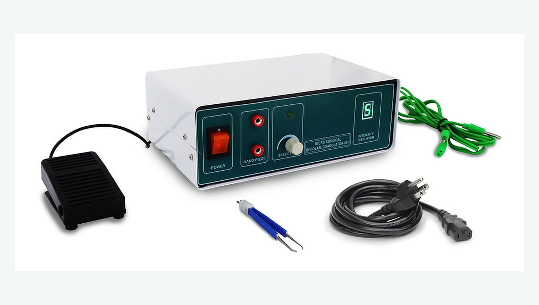
Provides precise coagulation of bleeding points during posterior-segment surgery.
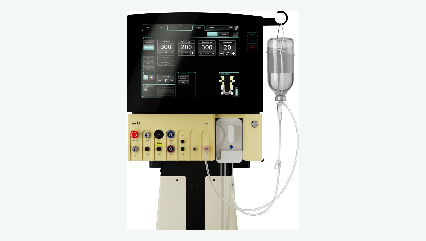
Supports silicone-oil injection and removal as well as fluid-air exchange for intraocular retinal tamponade.
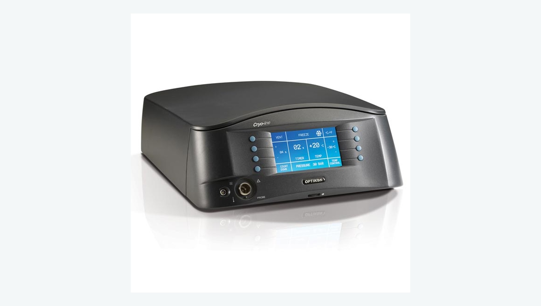
Uses a cryoprobe to create a chorioretinal adhesion or treat selected peripheral lesions.
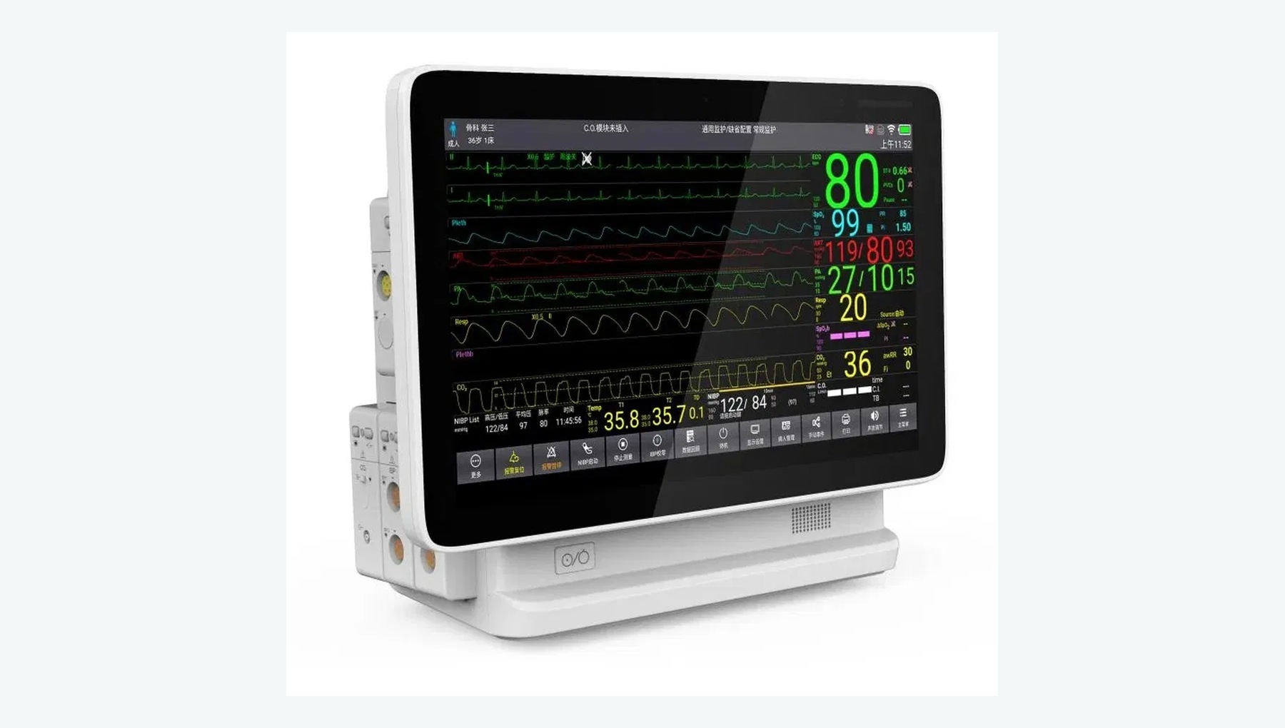
Monitors perioperative vital signs such as blood pressure, oxygen saturation and ECG.
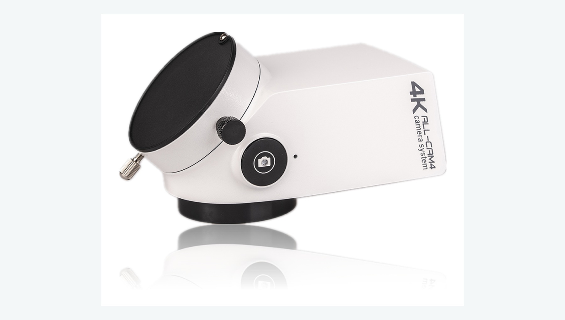
Records microsurgical procedures for case documentation, quality review and clinical training.
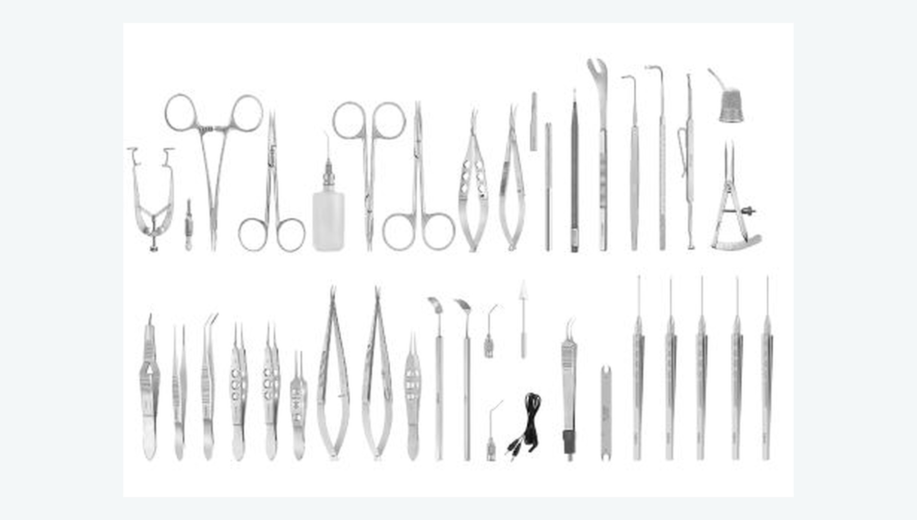
Supports sclerotomy creation, infusion, vitreous cutting, intraocular maneuvers and wound closure.
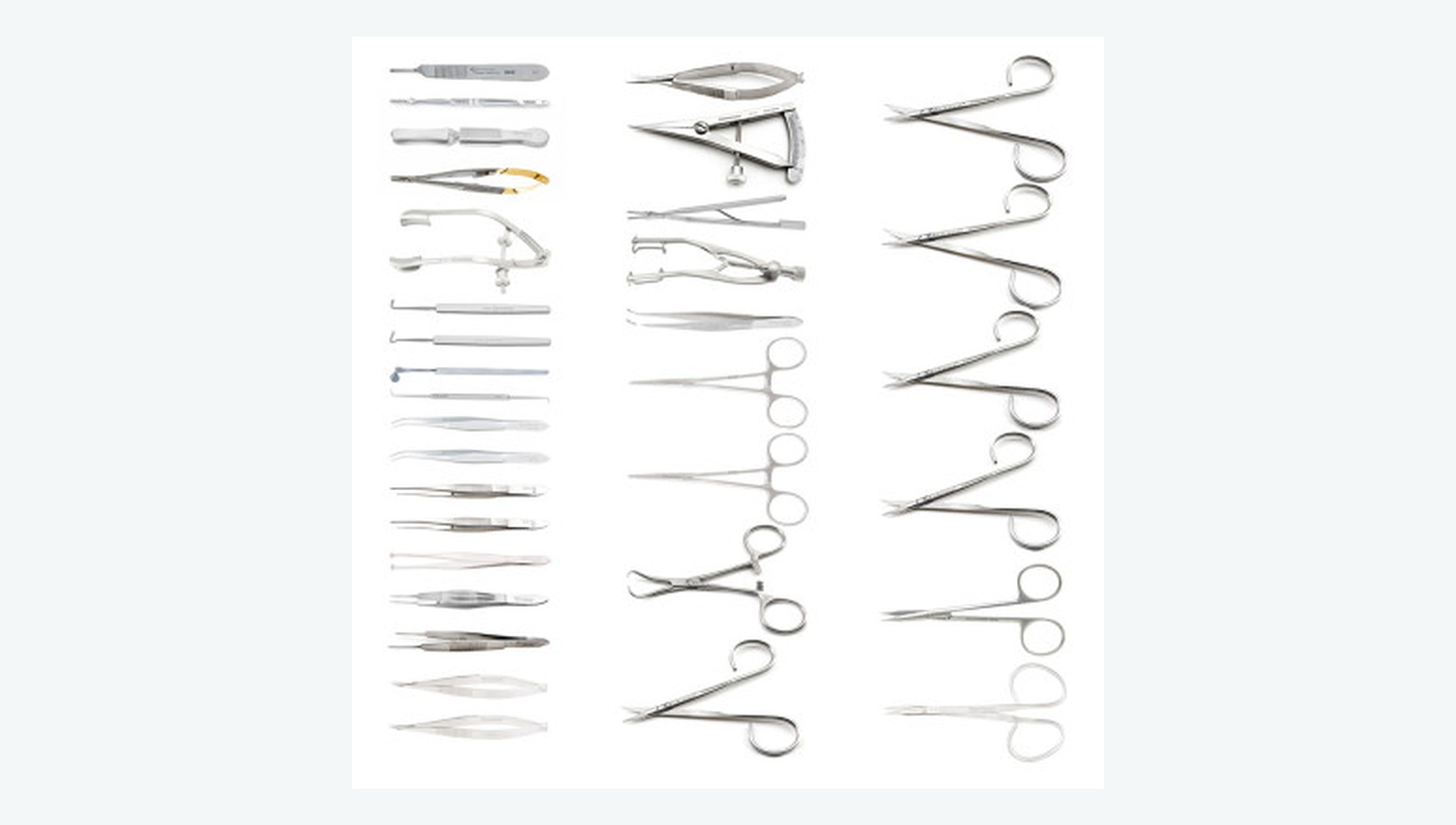
Supports tear localization, drainage, endolaser, fluid-air exchange and tamponade.
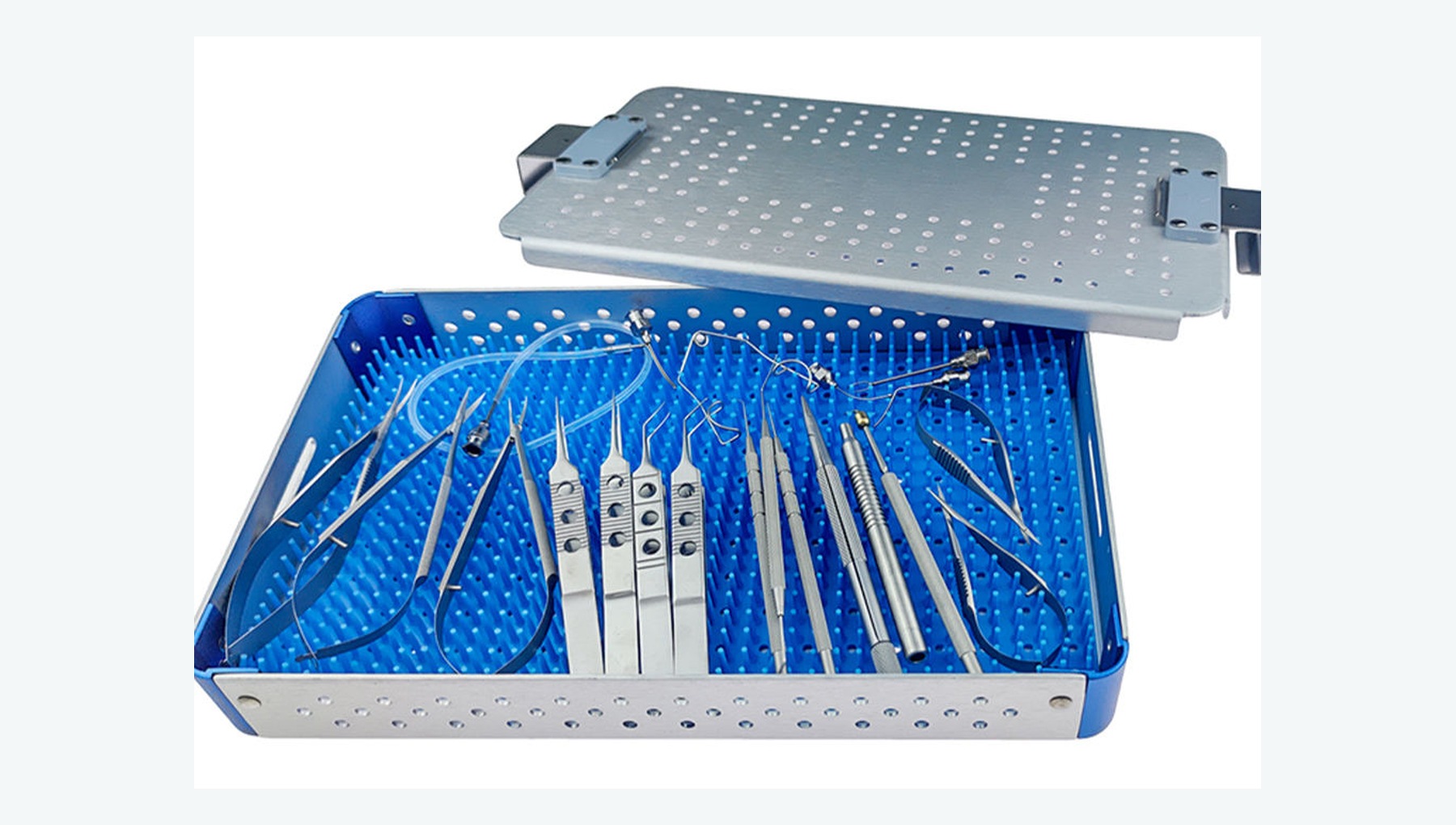
Supports extraocular muscle isolation, buckle placement, localization, drainage and fixation.
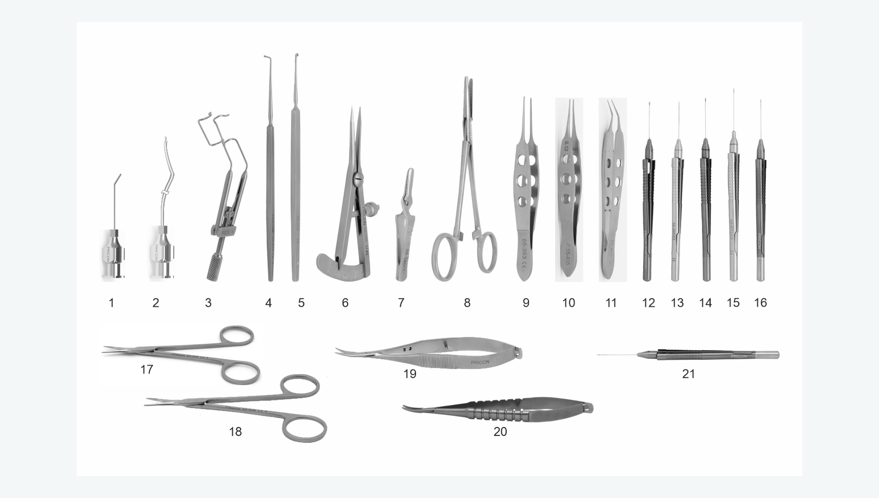
Supports fibrovascular membrane dissection, hemostasis, endolaser and complex vitreous surgery.
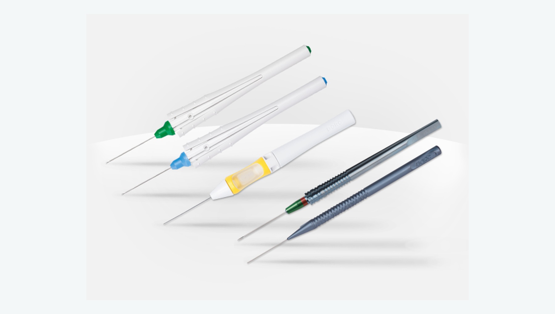
Supports staining and peeling of epiretinal and internal limiting membranes and other fine macular maneuvers.
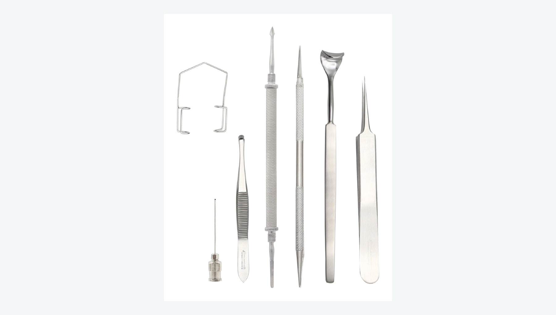
Supports posterior-segment trauma exploration, foreign-body localization and extraction, and related repair.
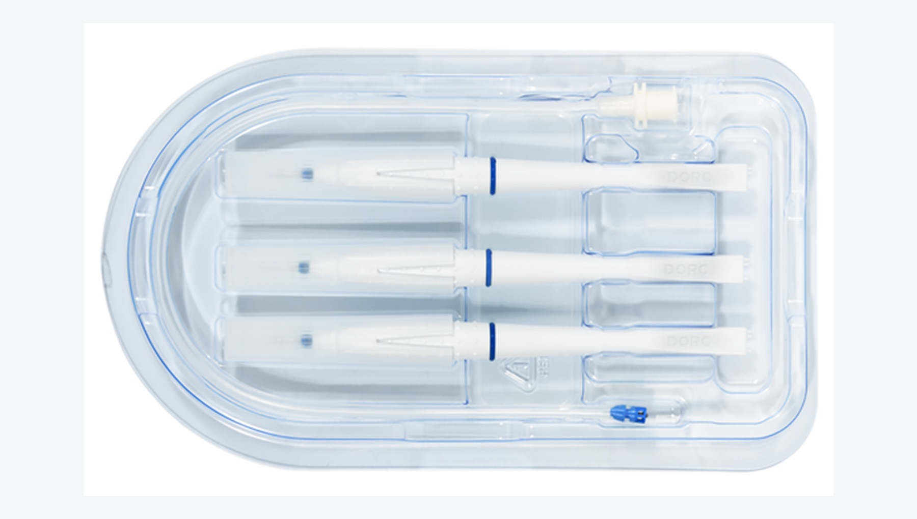
Supports aseptic intravitreal injection, ocular-surface preparation and post-procedure protection.
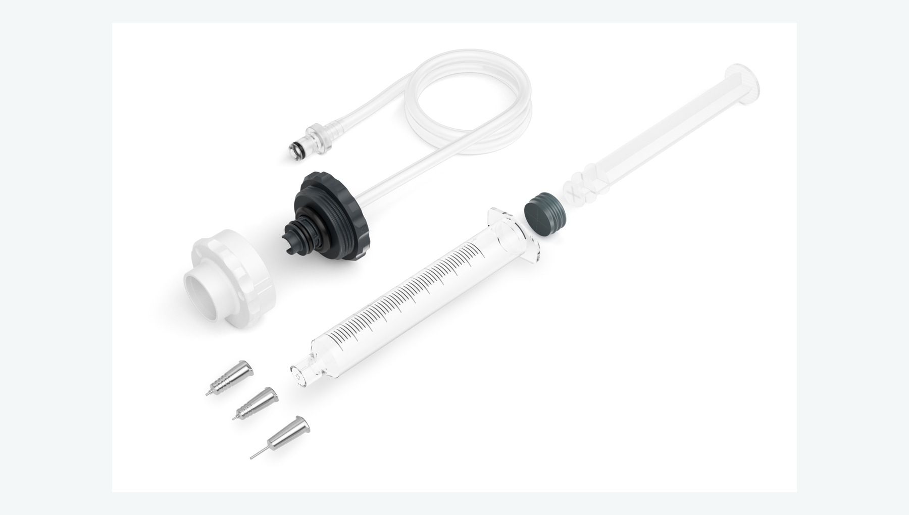
Supports silicone-oil or gas tamponade, silicone-oil removal and fluid-air exchange.
When your requirement includes room preparation or a complete department, connect the equipment brief with layout, engineering interfaces and team readiness.
Explore the related department or projectA concept connecting consultation, refraction, diagnostics, treatment and supporting spaces.
Explore plans & 3DLet’s plan the next step
Tell us what you want to build, improve or source. A short outline is enough to start.
 Chat with us